
|
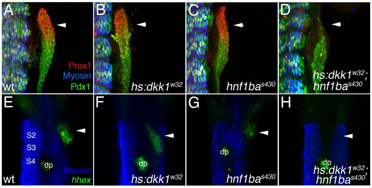
Fig. 4 Specification of hepatopancreas progenitors by Hnf1ba and Wnt signaling. (A-D) 3D rendering (lateral view showing somites for positional reference) of 36 hpf foregut endoderm in wild-type (A), Tg(hsp70l:dkk1-GFP)w32 (B), hnf1bas430 (C) and Tg(hsp70l:dkk1-GFP)w32; hnf1bas430 embryos (D) stained for Prox1, Pdx1 and myosin (somites). Relative to wild type, Prox1 foregut expression (arrowheads) is similar or mildly reduced in both Tg(hsp70l:dkk1-GFP)w32 and hnf1bas430 embryos. Prox1 expression is severely reduced or lost in Tg(hsp70l:dkk1-GFP)w32; hnf1bas430 embryos. (E-G) Double in situ hybridization and antibody staining (ventral view) for the early hepatopancreas progenitor marker hhex and myosin at 36 hpf. hhex is expressed (arrowheads) in the foregut endoderm and dorsal pancreas (dp) in wild type (E) but reduced in both the Tg(hsp70l:dkk1-GFP)w32 (F) and the hnf1bas430 (G) embryos. (H) hhex expression (arrowhead) is undetectable in the foregut endoderm of Tg(hsp70l:dkk1-GFP)w32; hnf1bas430 embryos.