
Fig. 2
- ID
- ZDB-IMAGE-130801-29
- Genes
- Publication
- Wang et al., 2013 - miR-34b regulates multiciliogenesis during organ formation in zebrafish
- All Figures
- Figures for Wang et al., 2013
|
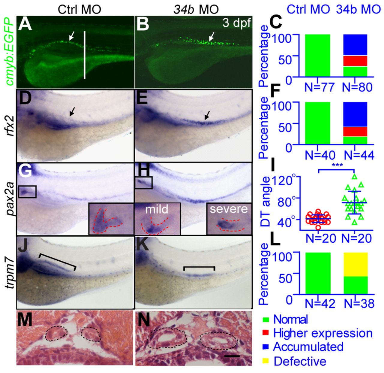
Fig. 2 Loss of miR-34b disrupts kidney morphogenesis. (A-F) In situ hybridization with the rfx2 probe and images of the Tg(cmyb:EGFP) embryo show that MCCs accumulate in the middle part of the pronephric duct (arrows) in miR-34b morphants at 3 dpf. C and F show the percentage of embryos with abnormal cmyb+ (shown in A,B) or rfx2+ (shown in D,E) MCCs. Embryos with accumulated MCCs (Accumulated) or higher expression levels of cmyb or rfx2 (Higher expression) were counted. (G-I) A magnified lateral view (with a small angle to dorsal) of a 3-dpf embryo after in situ hybridization with the pax2 probe shows that proximal tubule convolution is blocked at 3 dpf in miR-34b morphants. Examples of normal proximal tubule convolution (G) and of mild and severe defects (H) are illustrated in the insets. The angle between the kidney duct and tubule (DT angle) was measured. Each circle/triangle in I represents an embryo. Mean ± s.d. ***P<0.0001 (unpaired t-test). (J-L) Lateral view of a 3-dpf embryo after in situ hybridization with the trpm7 probe shows that transporting epithelial cells accumulate in the middle region (bracket) of the pronephric duct 3 dpf after miR-34b knockdown. L shows the percentage of embryos with abnormal distribution of trpm7+ transporting epithelial cells (Defective). (M,N) Hematoxylin and Eosin (H&E) staining on a transverse section (plane indicated by the white line in A) shows the enlarged tubule diameter (outlined) after miR-34b knockdown.