
|
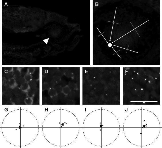
Fig. 8 Larval photoreceptor basal bodies are randomly positioned. A: Sagittal section through a 7-dpf Tg(-5actb2:cetn2-GFP)cu6 larvae shows the optic nerve (arrowhead) and larval retinal mosaic (arrow). B: Enlarged view of sectioned retina shows how to determine the orientation of cells relative to the optic nerve (circle). White arrows indicate the direction of the optic nerve, whereas red arrows indicate the 90° perpendicular angle. C,D: Immunostaining with zpr-1 (red) and GFP (yellow) antibodies identifies basal bodies in red-/green-sensitive cones. Blue-sensitive cones were negative for zpr-1 and 4C12 immunoreactivity (arrows). E: Inner and outer segments (autofluorescence from excitation with 488-nm light) and basal bodies (yellow) of UV-sensitive cones. F: Basal bodies (yellow) and 4C12 (red)-immunopositive rods. G-J: Mean angular positions of individual fields of red-/green-, blue-, and UV-sensitive cones and rods are plotted on circular graphs. Grand mean angular positions are shown as black squares. In all images, nuclei are counterstained with DAPI (blue). A magenta-green version of this figure is provided in the Supporting Information. Scale bar = 10 μm (C-F). [Color figure can be viewed in the online issue, which is available at wileyonlinelibrary.com.]