
|
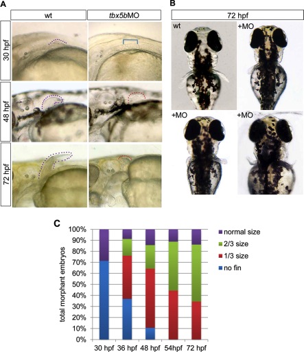
Fig. 8 Depletion of tbx5b delays pectoral fin initiation and reduces fin outgrowth. A: Lateral brightfield images of pectoral fin area taken of control or tbx5bMO-injected embryos; anterior is oriented to the left. Arrowheads denote the tip of the pectoral fin and are color-coded based on size (see B). B: Dorsal brightfield images of wild-type (wt) and tbx5bMO injected embryos at 72 hours postfertilization (hpf) illustrating the variability observed in pectoral fin morphology among morphant embryos. C: Quantification of pectoral fin size for tbx5bMO-injected embryos as development progresses. n = 21–46 embryos per time point. Dotted lines flank the fin bud tissue; bracket indicates area with missing fin.