
|
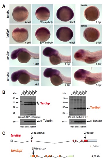
Fig. S1 Tardbp and Tardbpl are expressed throughout development and overview of zinc finger binding sites of the ZFN. (A) In situ hybridization with antisense probes specific for tardbp and tardbpl during early developmental stages of 4 cell to 5-dpf-old wild-type embryos. Labeling of 9-hpf-old embryos with a sense probe serves as a negative control (magnification: 10×). (B, Left) Western blot analysis with the Tardbp-specific antibody antiTDP-43 (Cosmo) reveals expression of Tardbp protein 1 dpf through 5 dpf. The asterisk marks an unspecific band. (Right) Western blot analysis with a Tardbpl-specific antibody (anti-Tardbpl 5F5-11) shows that Tardbpl is expressed 1 dpf through 5 dpf. Analysis with an anti–α-Tubulin specific antibody of the respective blots serves as a loading control. (C) Schematic representation of the genomic organization of tardbp (light blue boxes represent 5′ and 3′ UTR; red boxes represent coding exons) and tardbpl (light green boxes represent 5′ and 3′ UTR; orange boxes represent coding exons) locus. Arrows indicate genomic localization of the target sequence of respective ZFN sets.