
Fig. 2
|
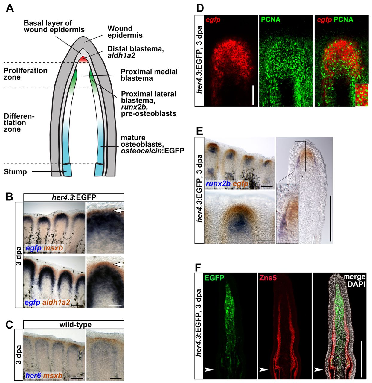
Fig. 2 Notch pathway activity is confined to the proliferative zone of the proximal medial blastema. (A) A longitudinal section of a regenerating fin during the outgrowth phase of regeneration (after 48 hpa) showing relevant anatomical structures and expression domains. (B) The her4.3:EGFP reporter (blue) is expressed proximally to msxb and aldh1a2 (red) at 3 dpa. (C) her6 (blue) is expressed proximal to msxb (red). (D) her4.3:EGFP is active in PCNA-positive cells. Confocal images of whole-mount regenerates stained for egfp transcripts and PCNA protein. (E) her4.3:EGFP (red) expression extends further distally than runx2b (blue), and is confined to the medial, runx2b-negative blastema. (F) Confocal images of longitudinal sections of her4.3:EGFP transgenic regenerates stained with Zns5 antibody (labeling all osteoblasts) and anti-GFP show no overlap. (B-E) Arrowheads indicate amputation plane. Scale bars: whole mounts, 200 μm; sections, 100 μm.