
Image
|
Figure Caption
Fig. 3
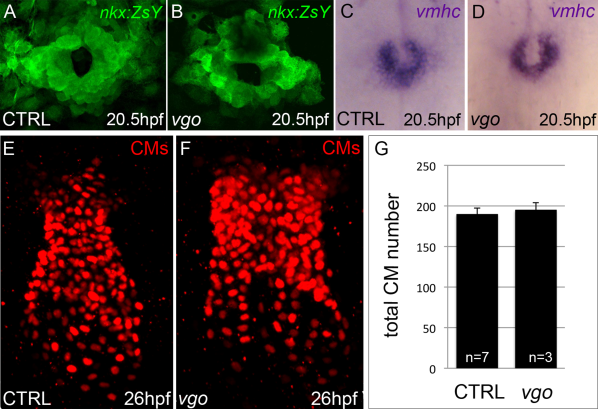
FIrst heart field development is unaffected in tbx1 mutants. A,B: Flattened confocal images of Tg(nkx2.5::ZsYellow) control (n=37; A) and vgo (n=14; B) embryos at 20.5hpf/23ss. C,D: Whole-mount in situ hybridization of vmhc at 20.5hpf/23ss in both control (n=16; C) and vgo (n=16; D) embryos. E,F: Flattened confocal images following immunofluorescence of cardiomyocyte nuclei comprising the linear heart tubes of 26 hpf control (E) and vgo (F) Tg(cmlc2:dsRed2-nuc) embryos. Dorsal view, anterior up in all images. G: Graph depicting the average number of CMs at 26 hpf in control and vgo embryos.
Figure Data
Acknowledgments
This image is the copyrighted work of the attributed author or publisher, and
ZFIN has permission only to display this image to its users.
Additional permissions should be obtained from the applicable author or publisher of the image.
Full text @ Dev. Dyn.