
IMAGE
Fig. 1, S2
- ID
- ZDB-IMAGE-130502-32
- Publication
- Saxena et al., 2013 - Sox10-dependent neural crest origin of olfactory microvillous neurons in zebrafish
- All Figures
- Figures for Saxena et al., 2013
Image
|
Figure Caption
Fig. 1, S2
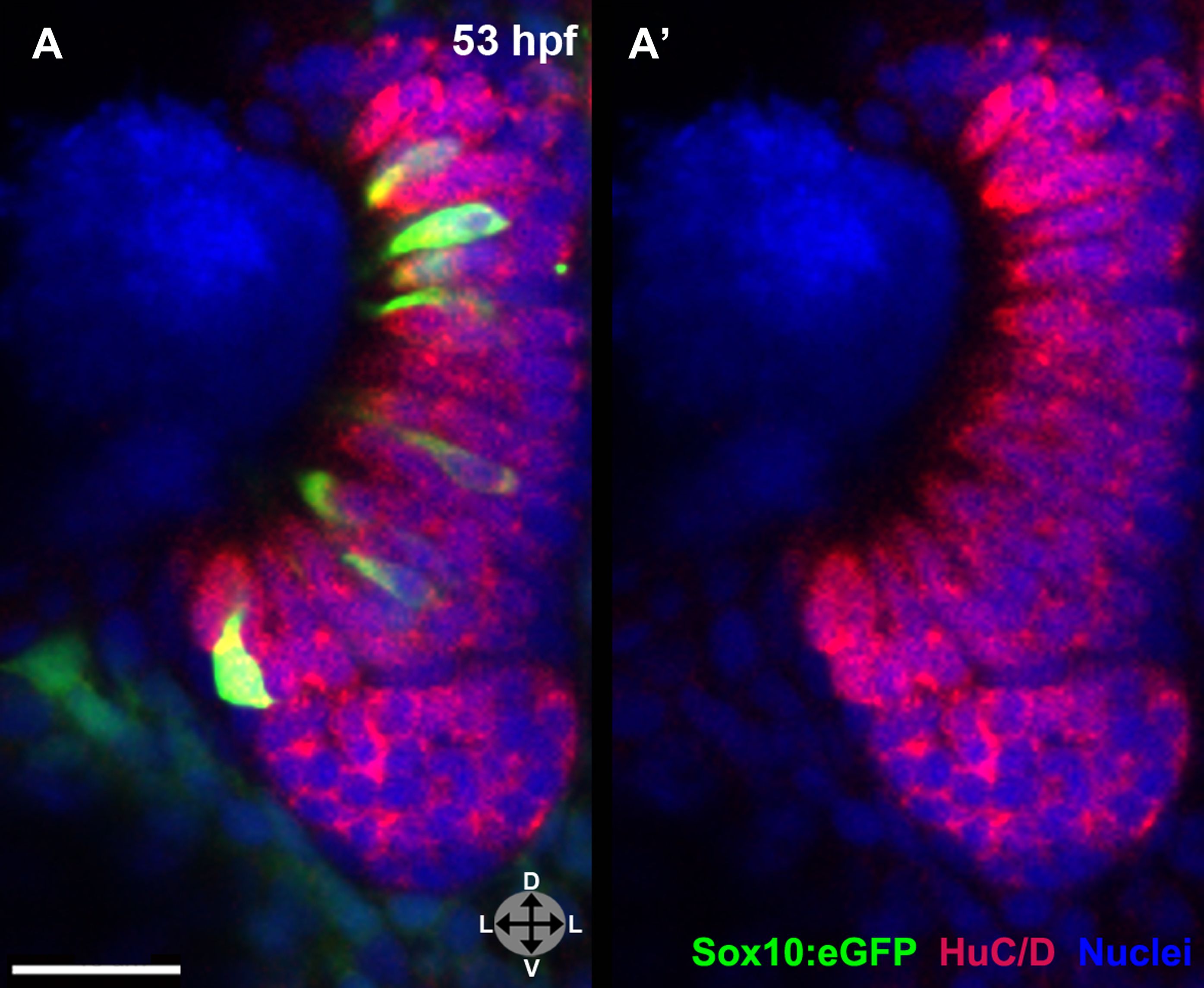
(A) and (A′) Sox10:eGFP+ microvillous neurons in fixed embryos stained with anti-GFP antibody colocalize with anti-HuC/D antibody staining at 53 hpf, confirming post-mitotic neuronal identity.
Sox10:eGFP: green; HuC/D: red; nuclear stain: blue. Orientation arrows: D: dorsal; V:ventral; L: lateral. z = 2.5 μm. Scale bar: 20 μm.
Acknowledgments
This image is the copyrighted work of the attributed author or publisher, and
ZFIN has permission only to display this image to its users.
Additional permissions should be obtained from the applicable author or publisher of the image.
Full text @ Elife