
IMAGE
Fig. 1, S1
- ID
- ZDB-IMAGE-130502-31
- Publication
- Saxena et al., 2013 - Sox10-dependent neural crest origin of olfactory microvillous neurons in zebrafish
- All Figures
- Figures for Saxena et al., 2013
Image
|
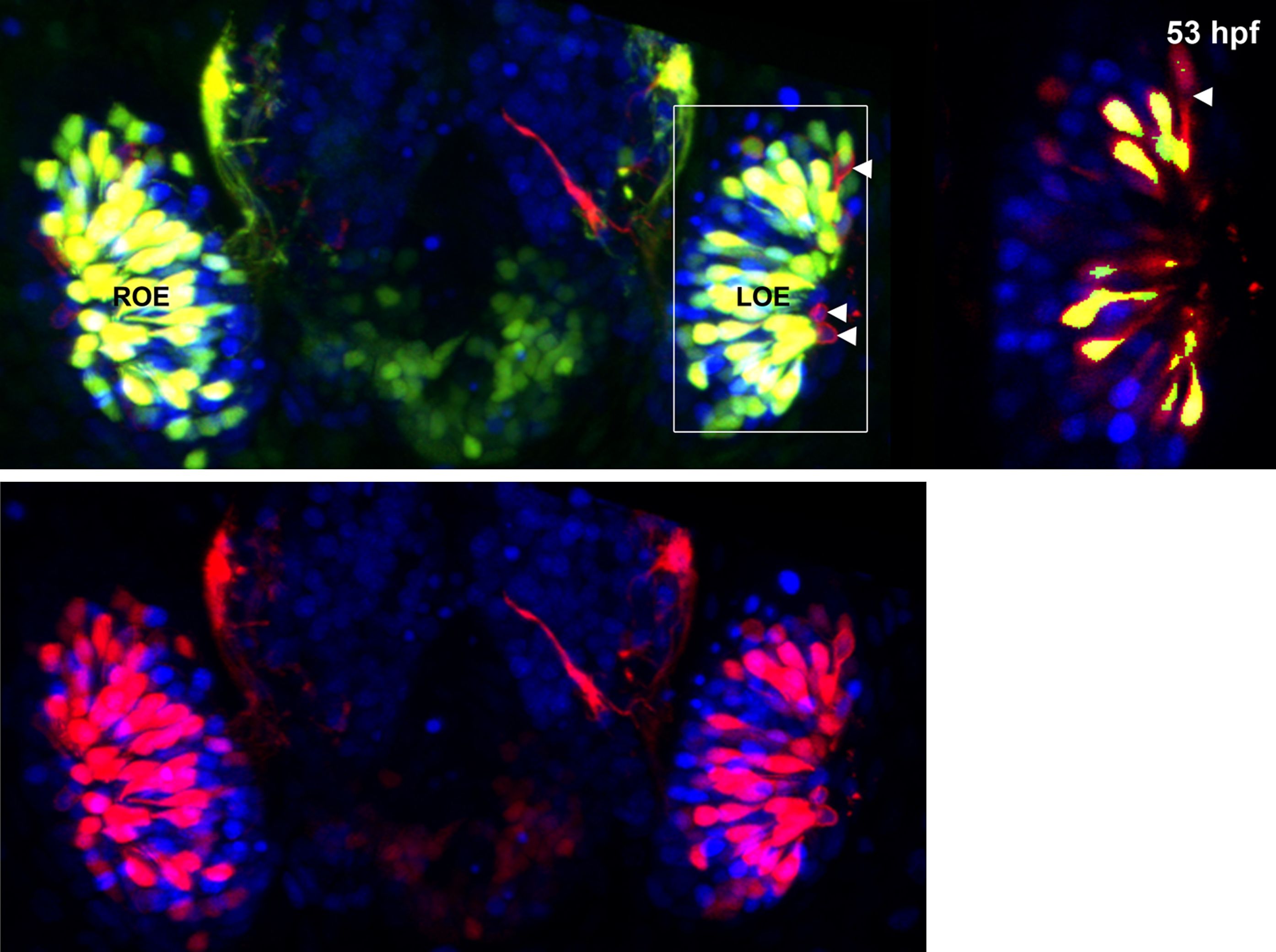
Figure Caption
Fig. 1, S1 Panel (K) from Figure 1 with Sox10:eGFP (green) channel removed (bottom) to better illustrate the large population of ciliated neurons present basally that are not Sox10:eGFP+ but are Ngn1:nRFP+ (blue).
High magnification inset (right) has Sox10:eGFP signal artificially underexposed and decreased to better view the colocalization with membrane TRPC2:Venus expression.
Acknowledgments
This image is the copyrighted work of the attributed author or publisher, and
ZFIN has permission only to display this image to its users.
Additional permissions should be obtained from the applicable author or publisher of the image.
Full text @ Elife