
|
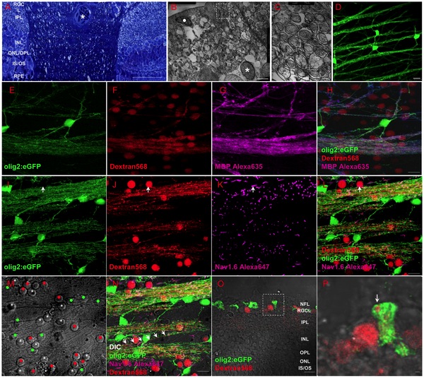
Fig. 4 Oligodendrocytes exist within retina of adult zebrafish.
(A) Toluidin blue staining shows myelin structure through the lamina cribrosa into the retina. (B-C) TEM images show that the axons of RGCs are specially and loosely wrapped by myelin. (D) Olig2:eGFP cells in retina have perfect structure of oligodendrocyte cells that project several processes to vicinal axons in Tg(olig2:eGFP) transgenic line. (E-H) Combination of Dextran568 labeled RGCs and MBP staining show olig2+ eGFP expression on mature oligodendrocytes in NFL. Notice that only the myelin ensheathed axons are MBP positive. (I-N) With Ranvier node on myelin, oligodendrocytes in zebrafish retina are in mature state. Arrow in (I-L) points out an axon hillock near RGC soma, and it should be noticed that olig2+ process dose not wrap this area. Red dots in (M) indicate RGCs, green dots are olig2+ cells and * indicates non-labeled cells, a row of arrows in (N) show an axon is wrapped by olig2+ process. (O) Crossing section indicates that axon retrogradely labeled by Dextran568 is wrapped by olig2+ eGFP. (P) Details of the white square in (O). Scale bar: 50 μm (A); 2 μm (B); 200 nm (C); 10 μm (D-O); 1 μm (P).