
|
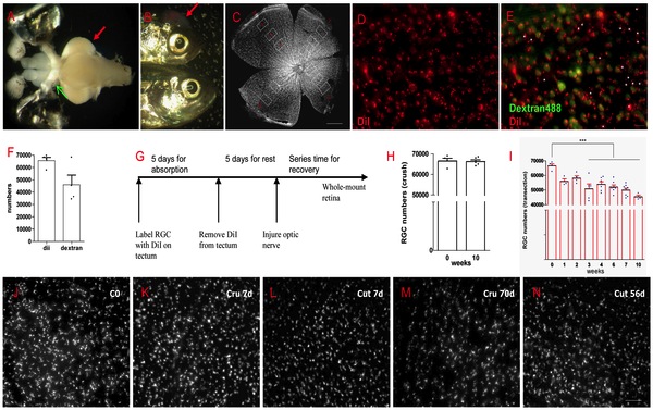
Fig. 1 Most RGCs survived after ONI.
(A) Two methods were used to label RGCs, the red arrow indicates DiI placed on tectum, the green arrow indicates Dextran 488 at optic nerve stump. (B) The red arrow shows lightcuring bond on the skull to prevent DiI lost. (C) The sampling of RGCs counting in the entire retina. (D) An example of DiI labeled RGCs. (E) Merge DiI and Dextran labeled RGCs in the same field. Asterisks indicate cells labeled by DiI but not by Dextran488. (F) Though the number of labeled cells has no significant difference (p>0.05) between DiI and Dextran, retrograde labeling with DiI is more reliable. (G) A scheme of DiI labeling in RGC survival experiment. (H) Almost all RGCs survived in ONC model after 10 weeks. (I) Significant RGCs lost began at 3 weeks (about 20% lost) but over 70% of RGCs were still alive at 10 weeks after ONT, p<0.001. (J-N) Representative pictures show RGCs at different time in ONC or ONT model. * indicates p<0.05, *** is p<0.001. Scale bar: 200 μm (C); 10 μm (E); 30 μm (J-N).