
|
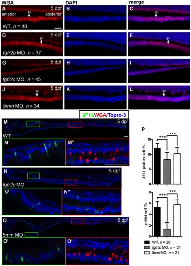
Fig. 3
The secretory cell differentiation of fgfr2c morphants.
WGA staining was performed on 5 dpf (A–C) WT embryos, (D–F) fgfr2b, (G–I) fgfr2c, and (J–L) fgfr2c-5 mm morphants. White arrows indicated goblet cell (gc). Double labelling using 2F11 antibody and WGA in (M) WT embryos, (N) fgfr2c morphants, and (O) fgfr2c-5 mm morphants. The magnified image shows (M′–O′) enteroendocrine cells and (M′ ′–O′ ′) goblet cells. Topro-3 was used for nuclear counter staining (blue). (P) The bar charts show the percentages of 2F11 or WGA positive cells. All images were lateral view with anterior at left and posterior at right. Error bars indicate SD. Scale bars = 50 μm.