
|
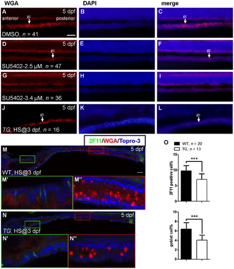
Fig. 1
Effects on cell differentiation after inhibition of Fgf signaling.
Five dpf embryos were stained with WGA after incubation in (A–C) DMSO, (D–F) SU5402 (2.5 μM), and (G–I) SU5402 (3.4 μM) at 3 dpf. (J–L) Three dpf Tg(hsp70l:dnfgfr1-EGFP) embryos were heat treated, then stained with WGA at 5 dpf. White arrow indicated the goblet cell (gc). DAPI nuclear counter stain showed the tissue structure. (M) Heat-treated WT embryos were used as controls. (N) Heat shocked transgenic embryos were double labeled with WGA and 2F11 antibody at 5 dpf. (M′ ,N′) The magnified image shows enteroendocrine and (M′ ′,N′ ′) goblet cells. (O) The bar charts show the percentage of 2F11 or WGA positive cells. All images were lateral view with anterior at left and posterior at right. Error bars indicate SD. Scale bars = 50 μm.