
|
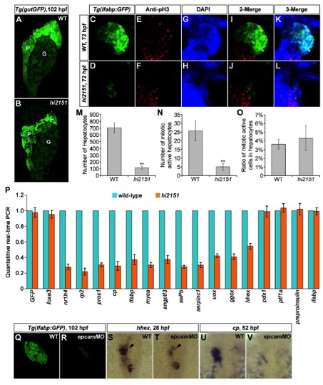
Fig. S2
Defective Liver Development in hi2151 Mutants or epcam Morphants. related to Figure 1
(A and B) Liver phenotypes in hi2151 mutants at 102 hpf under the Tg(gutGFP) transgenic background. Note that the pancreas and gut remain relatively normal. L, liver; P, pancreas; G, gut.
(C and D) Liver phenotypes in hi2151 mutants at 72 hpf under the Tg(lfabp:GFP) background.
(E and F) Antibodies against phosphorylated histone 3 (pH3) mark the mitotic active cells in the liver.
(G and H) DAPI stainings mark the nuclei of liver cells.
(I) Merge of (C) and (E) to indicate the mitotic active hepatocytes.
(J) Merge of (D) and (F) to indicate the mitotic active hepatocytes.
(K) Merge of (C), (E), and (G).
(L) Merge of (D), (F), and (H).
(M) In contrast to the wild type, the number of hepatocytes per embryo at 72 hpf is significantly reduced in hi2151 mutants. n=5, **, p<0.01, Student’s t test.
(N) In contrast to the wild type, the number of mitotic active hepatocytes per embryo at 72 hpf is significantly reduced in hi2151 mutants. n=5, **, p<0.01, Student’s t test.
(O) In contrast to the wild type, the ratio of mitotic active hepatocytes at 72 hpf remains in hi2151 mutants. n=5, p>0.05, Student’s t test.
(P) Endodermal cells are sorted from beheaded wild-type and hi2151 mutant embryos under the Tg(sox17:GFP) background at 52 hpf. Transcriptions of twelve hepatic genes (www.zfin.org, nr1h4, rp2, prox1, cp, lfabp, myca, angptl3, sePb, serpinc1, uox, ggcx, and hhex), GFP, the endodermal marker foxa3, the pancreatic/intestinal markers pdx1, ptf1a, preproinsulin, and ifabp are analyzed by quantatitive real-time PCR.
(Q and R) Liver phenotypes in epcam morphants at 102 hpf as shown under the Tg(lfabp:GFP) background.
(S–V) Expressions of hhex in the hepatic endoderm at 28 hpf (S and T, arrowheads) and cp at 52 hpf (U and V).
Error bars represent SD.
Reprinted from Developmental Cell, 24(5), Lu, H., Ma, J., Yang, Y., Shi, W., and Luo, L., EpCAM Is an Endoderm-Specific Wnt Derepressor that Licenses Hepatic Development, 543-553, Copyright (2013) with permission from Elsevier. Full text @ Dev. Cell