
|
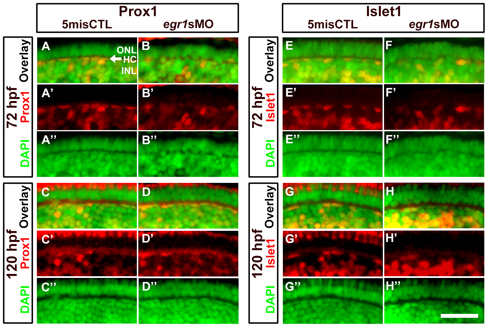
Fig. 6 Immunohistochemical analysis of the HCs in the Egr1-morphant retinas.
A magnified view of the immunostaining results of HCs in the controls (5misCTLMO) and Egr1 morphants (egr1sMO) with Prox1 (A-D) and Islet1 (E-H) at 72 and 120 hpf. These selected regions correspond to the white boxes as shown in Figure 4. Prox1+ and Islet1+ cells are shown in red, while the DAPI nuclei counterstain is shown in green. The location of HCs is indicated by an arrow in A. See text, Table 1 and 2 for further discussion. In all pictures, the apical retina is to the top and dorsal is to the left. HC: horizontal cells; INL: inner nuclear layer; ONL: outer nuclear layer. Scale bar = 50 µm.