
|
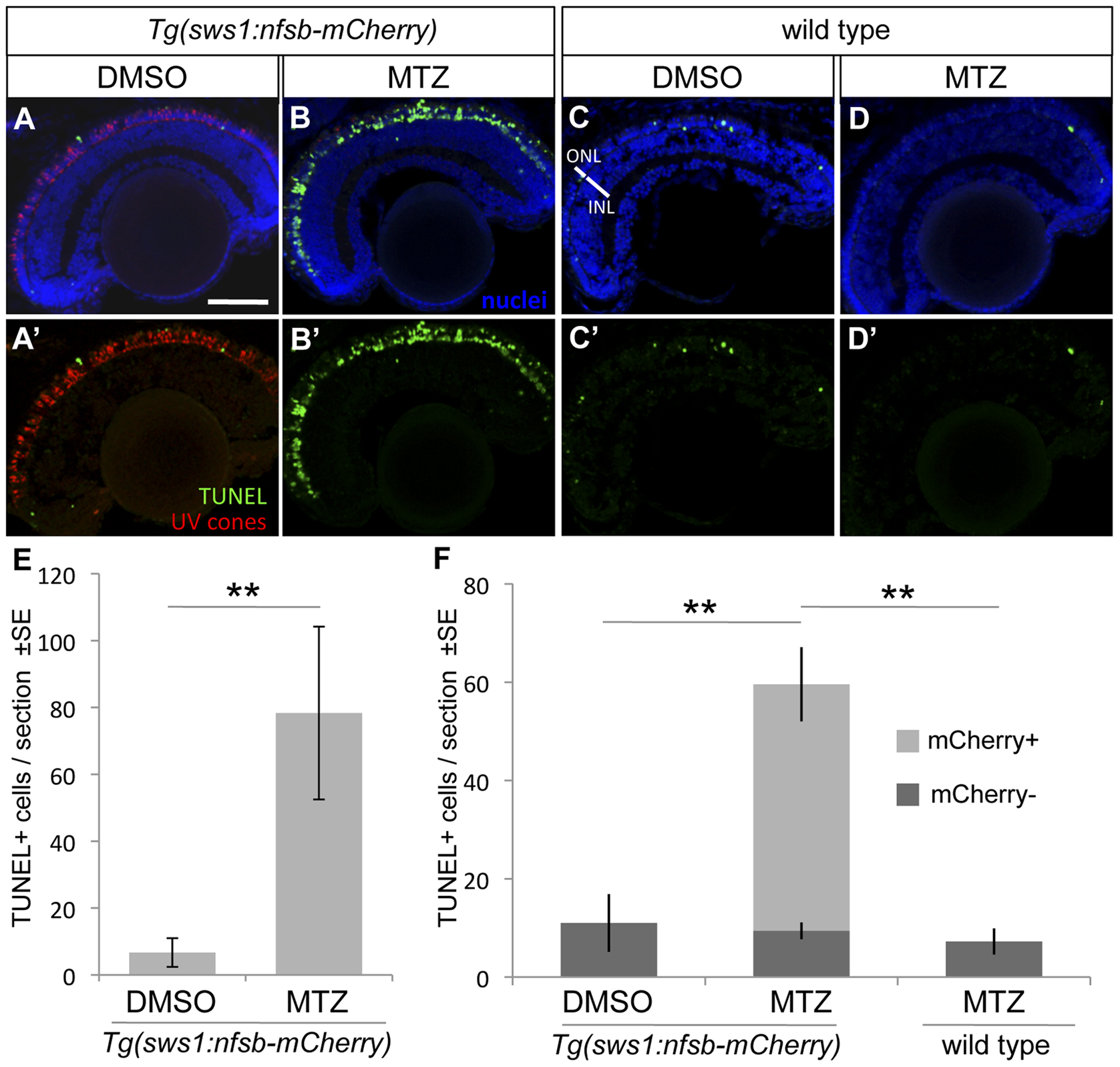
Fig. 4 UV cone photoreceptor death induced by prodrug application to transgenic fish.
An abundance of apoptotic cells were detected in the ONL of the retina of Tg(SWS1:Gal4-VP16)ua3016;Tg(UAS-E1b:NfsB-mCherry)c264 fish treated with MTZ for 48 hrs (B, B′) compared to various controls. Very little apoptosis was found in the ONL of Tg(SWS1:Gal4-VP16)ua3016;Tg(UAS-E1b:NfsB-mCherry)c264 fish treated with a control DMSO solution (A, A′). Non-transgenic siblings treated with either DMSO or MTZ (C or D, respectively) for 48 hrs also possessed few apoptotic cells. E. The number of TUNEL+ cells in retinal sections equivalent to A and B were quantified, showing an order of magnitude increase upon MTZ prodrug application (**p<0.001, MTZ-treated n = 11, DMSO treated n = 15). F. A repetition of this labelling to discern the bystander effect, i.e. compare the number of TUNEL+ cells that do not express nfsb-mCherry as compared to basal levels in normally developing fish. Again the total number of TUNEL+ cells is increased when the transgenic fish are treated with prodrug MTZ rather than DMSO vehicle control (compare total height of light+dark bars, MTZ-treated n = 7). This contrasts the abundance of TUNEL+ cells without mCherry, which is not increased relative to DMSO control fish nor to wildtype fish receiving MTZ (dark grey bars, p = 0.84 and p = 0.927, respectively wherein n = 4 or 3, with means comparable to panel E). Scale bar = 50 µm.