
|
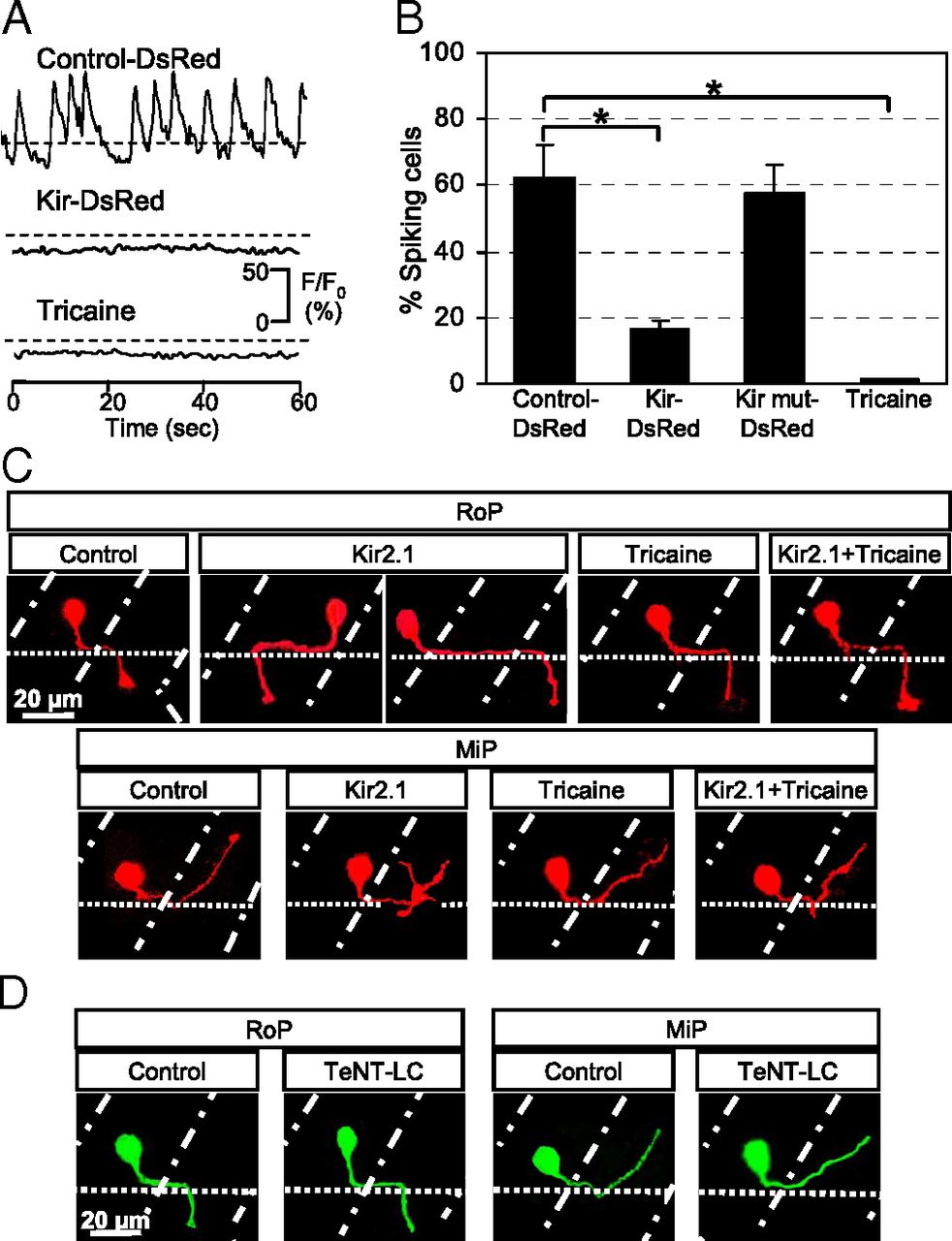
Fig. 6 Suppression of Ca2+ spiking activity in single PMN leads to errors in axon pathfinding. (A) Ca2+ spikes are blocked by stochastic expression of hKir2.1 or exposure to 0.02% tricaine for 15 min. n = 20 for each condition. (B) Percentage of PMN exhibiting Ca2+ spiking activity at 19 hpf. n = 20 for each group; values are means ± SEM. *P < 0.0005 compared with control. (C) RoP and MiP neurons expressing hKir2.1 bypass the exit point or project rostrally and exhibit extra branching. Neither tricaine nor Kir2.1 + tricaine leads to errors in axon pathfinding. Control, embryos injected with Hb9:Gal4 and UAS:DsRed plasmids; Kir2.1, embryos injected with Hb9:Gal4 and UAS:DsRed::UAS:hKir2.1 plasmids; Tricaine, embryos raised in the presence of 0.02% tricaine; Kir2.1 + Tricaine, embryos expressing hKir2.1 and raised in the presence of tricaine. (D) Tetanus toxin expression does not cause pathfinding errors. Control, embryos injected with Hb9:Gal4 and UAS:eGFP plasmids; TeNT-LC, embryos injected with Hb9:Gal4 and UAS:TeNT-LC:eGFP plasmids. (C and D) Dorsal is to the top and rostral is to the left. Dot-dash lines mark lateral edges of the myotomes; dotted lines mark the ventral edge of the spinal cord. n ≥ 30 cells from ≥30 24-hpf embryos for each group.