
|
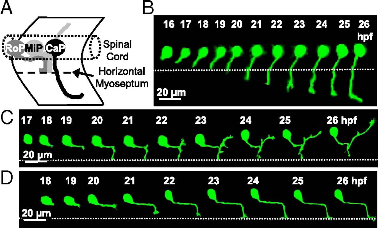
Fig. 1 PMN axon pathfinding in the developing zebrafish embryo. (A) Schematic lateral view of the three PMN (CaP, MiP, and RoP) showing their axonal trajectories at 24 hpf. Anterior to the left and dorsal to the top. (B–D) Time-lapse analysis of axon outgrowth of a single CaP (B), MiP (C), or RoP (D) expressing eGFP in WT embryos. Each neuron was imaged at 20-min intervals for 10 h. The dotted line indicates the HMS. CaP is present at 16 hpf and begins to extend a process by 17 hpf. At 19 hpf, coincident with its arrival at the HMS, the CaP axon pauses, resuming its outgrowth by 21 hpf. MiP starts axonogenesis by 18 hpf and is followed by RoP at 19 hpf. n = 10 neurons of each class in 10 embryos.