
|
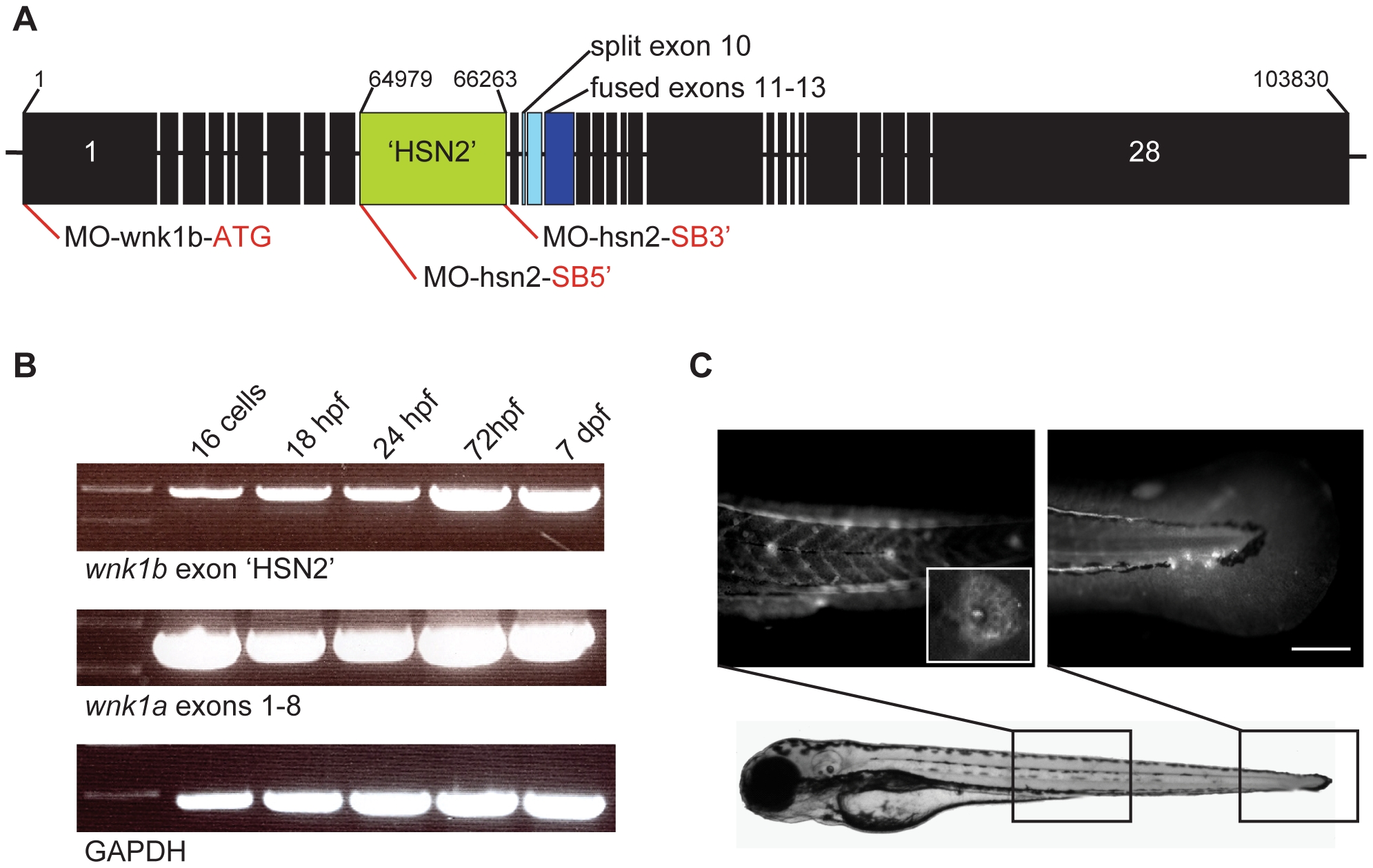
Fig. 1
Expression of the WNK1 kinase in zebrafish embryos.
A) Structure of the zebrafish WNK1 ortholog which conserved the HSN2 exon, wnk1b. The split exon 10 and fused exons 11–13 have been indicated on the sequence, respectively in pale blue and dark blue, as well as antisense morpholino oligonucleotide targets (red lines). B) Both copies of the zebrafish WNK1 ortholog, wnk1a and wnk1b are expressed early at the 16 cells stage and persist at 18, 24 and 72hours post-fertilization (hpf) and until 7 days post-fertilization (dpf). The RT-PCR was done using primers set in the HSN2 exon for wnk1b, targeting a sequence spanning exons 1–8 for wnk1a and using as control the housekeeping gene GAPDH. C) Zebrafish WNK1/HSN2 from the wnk1b gene was detected in the neuromasts of the posterior lateral line by whole-mount immunohistochemistry using an anti-HSN2 antibody. The inset shows a closer view of a stained neuromast. Scale bar: 100μm.