
Fig. 4
- ID
- ZDB-IMAGE-130123-61
- Genes
- Publication
- Bhat et al., 2013 - A gene network that coordinates preplacodal competence and neural crest specification in zebrafish
- All Figures
- Figures for Bhat et al., 2013
|
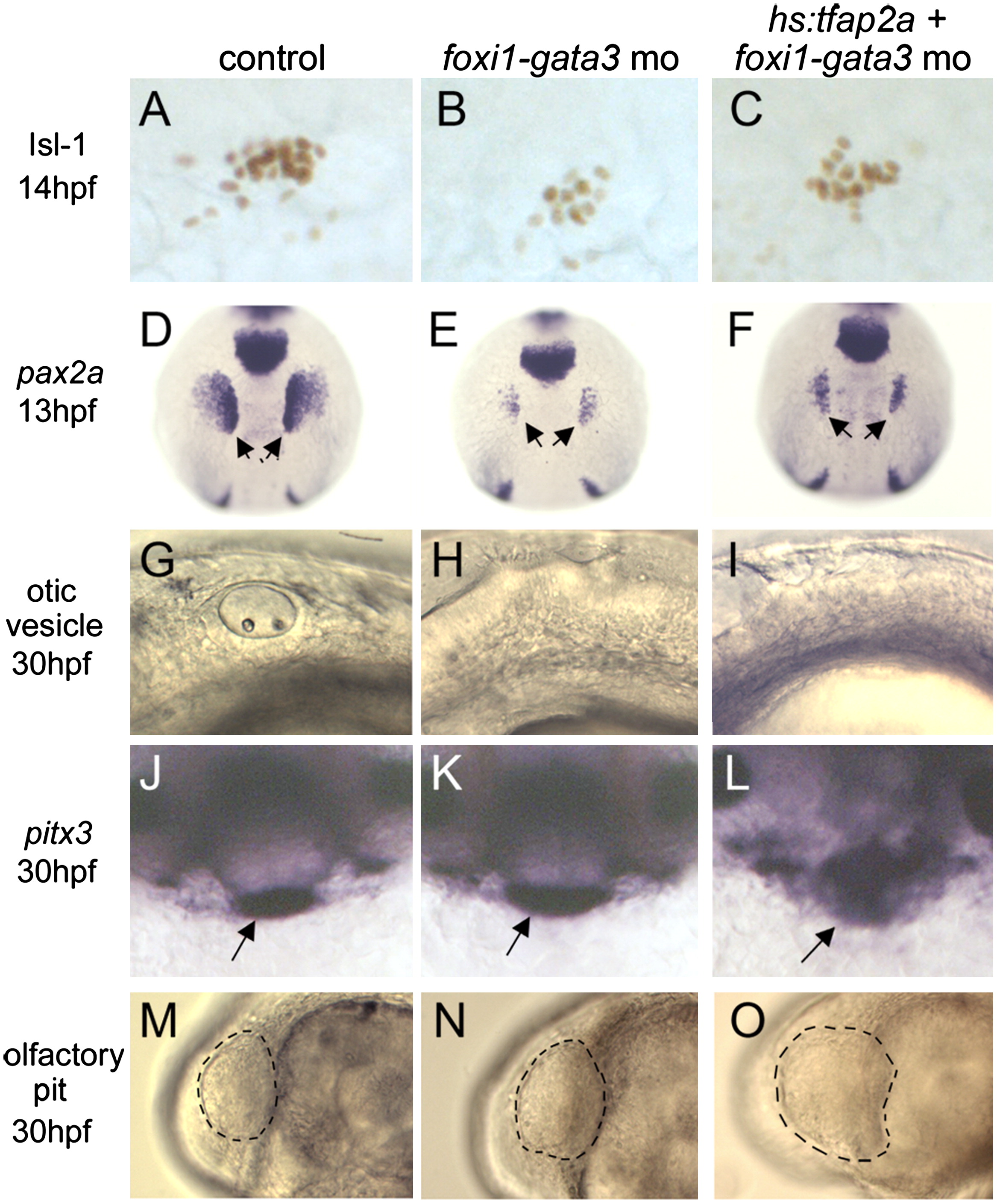
Fig. 4 Unique requirements for foxi1 and gata3 in posterior placodes. Markers of cranial placodes and placodal derivatives, including Isl1 in the trigeminal placode at 14 hpf (A–C), pax2a in the otic/epibranchial placodes at 13 hpf (D-F, arrows), otic vesicle at 30 hpf (G-I), pitx3 in the anterior pituitary at 30 hpf (J-L, arrows) and the olfactory pit at 30 hpf (M–O, outlined) in control embryos (A, D, G, J and M), foxi1-gata3 morphants (B, E, H, K and N), and foxi1-gata3 morphants in which hs:tfap2a was activated at 7 hpf (C, F, I, L and O). Knockdown of foxi1 and gata3 reduces the size of the trigeminal, otic and epibranchial placodes, but anterior placodes develop normally. Activation of hs:tfap2a does not rescue development of posterior placodes in foxi1-gata3 morphants, and the transgene still enlarges anterior placodes despite knockdown of foxi1 and gata3. Images show lateral views with anterior to the right (A-C, G-I, M-O), dorsal views with anterior up (D-F), and facial views of the front of the head (J-L).
Reprinted from Developmental Biology, 373(1), Bhat, N., Kwon, H.J., and Riley, B.B., A gene network that coordinates preplacodal competence and neural crest specification in zebrafish, 107-117, Copyright (2013) with permission from Elsevier. Full text @ Dev. Biol.