
|
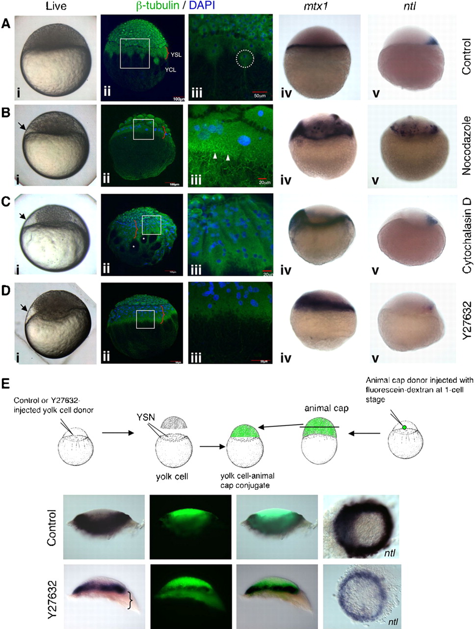
Fig. 3 Disruption of cytoskeletal elements induces formation of enlarged YSL.
(A) Control, (B) nocodazole-treated, (C) cytochalasin D-treated, and (D) Y27632-injected embryos between oblong to sphere stages. Arrows in Bi–Di indicate enlarged YSL. Curly brackets in Aii–Dii indicate the width of the YSL microtubule belt. (Aiii–Diii) High magnification images of boxes in Aii–Dii respectively. (Aii,Aiii) Microtubule distribution in a control embryo. A large aster in the YSL (stippled circle, Aiii). (Bii,Biii) Disorganized microtubules in the YSL and YCL, with small asters in the enlarged YSL (arrowheads, Biii). (Cii,Ciii) Gaps in the YCL (Ciii, asterisks). (Dii,Diii) Dense YSL and sparse YCL microtubule arrays. The enlarged syncytia express mtx1 (Biv–Div), and induces ntl in the overlying blastoderm (Bv–Dv). (E) Mesoderm induction assay. The enlarged syncytium (curly bracket) induced ntl in the fluorescein-labelled animal cap.