
|
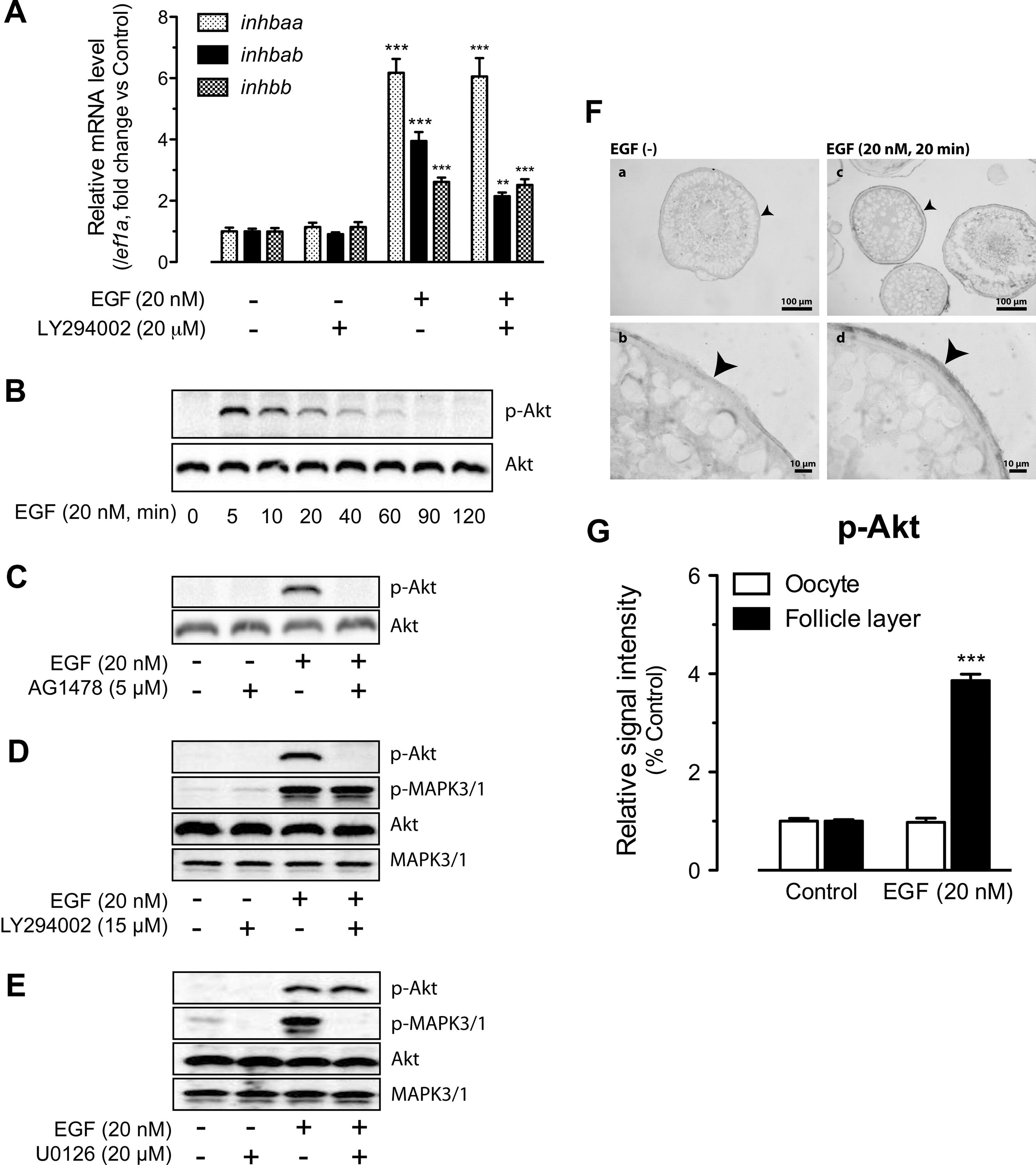
Fig. 3
Involvement of PI3K/Akt in EGFR signaling and EGF-stimulated activin subunit expression. (A) Effects of LY294002 (PI3K inhibitor) on EGF-stimulated expression of inhbaa, inhbab and inhbb in cultured follicle cells. The cells were treated with EGF at 20 nM for 2 h. The expression values are the mean ± SEM (n = 3–4). **P < 0.01; ***P < 0.001 vs. respective control of each gene. The graph is representative of four independent experiments. (B) Time course of EGF-induced Akt phosphorylation (p-Akt) in the follicle cells. (C) Effects of EGFR inhibitor AG1478 on basal and EGF-induced Akt phosphorylation. (D) Effects of PI3K inhibitor LY294002 on Akt and MAPK3/1 phosphorylation. (E) Effects of MEK1/2 inhibitor U0126 on Akt and MAPK3/1 phosphorylation. The follicle cells were pre-treated with the inhibitors for 30 min before treatment with EGF for 5 min. (F) Immunohistochemical staining for EGF-induced Akt phosphorylation in intact follicles. The follicles were treated with (20 nM) or without EGF for 20 min before sampling for fixation, sectioning and IHC staining. No signal was detected in the control follicles without EGF treatment (a and b), whereas signal of Akt phosphorylation was detected in the somatic follicle layer (arrow head) but not the oocyte (c and d). (G) Semi-quantitative measurement of immunohistological staining for p-Akt in the follicle layer and oocyte compartments of intact follicles. p-Akt level increased in the follicle layer after EGF treatment. The values are the mean ± SEM (n = 4). ***P < 0.001 vs. control.
Reprinted from Molecular and Cellular Endocrinology, 361(1-2), Chung, C.K., and Ge, W., Epidermal growth factor differentially regulates activin subunits in the zebrafish ovarian follicle cells via diverse signaling pathways, 133-142, Copyright (2012) with permission from Elsevier. Full text @ Mol. Cell. Endocrinol.