
|
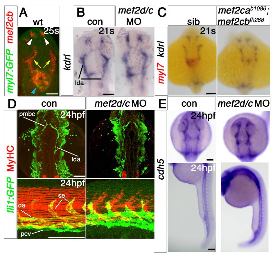
Fig. S4
Endothelial markers are little affected by Mef2 knockdown.
In situ mRNA hybridisation for mef2cb (A), kdrl (B and C), myl7 (C, red) and cdh5 (E) or immunodetection of MyHC and GFP (D) in wild type or Tg(fli1:GFP) control or injected with mef2d/c MO (except C; mef2cab1086;mef2cbfh288 and sibling embryos). A. Mef2cb mRNA detected in heart (blue arrowhead), telencephalon (white arrowheads), and head vasculature (yellow arrows). B and C. Kdrl (flk-1) expression in mef2d/c morphant and double mef2ca;mef2cb mutant embryos at 21 ss is not changed compare with control and sibling embryos, respectively. D. Expression of endothelial marker fli1:GFP at 24 hpf is similar in control and mef2d/c morphants in the heart and head region (upper panels), but some defects in intersomitic vessels of morphants (lower panels) parallel the somitic muscle MyHC phenotype previously described (Hinits and Hughes, 2007). E. Mild upregulation of cdh5 mRNA in head and heart region (upper panels) and in the trunk region (lower panels) at 24 hpf. lda, lateral dorsal aorta; pmbc, primordial midbrain channel; da, dorsal aorta; pcv, posterior cardinal vein; se, intersegmental vessel. Scale = 100 μm.
Reprinted from Developmental Biology, 369(2), Hinits, Y., Pan, L., Walker, C., Dowd, J., Moens, C.B., and Hughes, S.M., Zebrafish Mef2ca and Mef2cb are essential for both first and second heart field cardiomyocyte differentiation, 199-210, Copyright (2012) with permission from Elsevier. Full text @ Dev. Biol.