
|
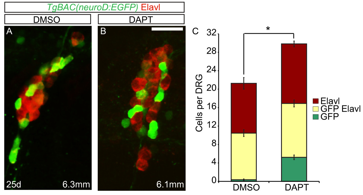
Fig. 7 Excess neurons differentiate in late larval dorsal root ganglia (DRG) following Notch inhibition. (A-B) Confocal images of TgBAC(neurod:EGFP) transgenic fish treated with DMSO or DAPT between 20 and 25 dpf. DRG neurons were labeled with anti-GFP antibody (green) and Elavl (red). At 25 dpf, the DRG of a 6.3 mm DMSO-treated fish (A) and a 6.1 mm DAPT-treated fish (B) contain neurons that are labeled with Elavl, GFP/Elavl or GFP antibody alone. (C) Quantification of cells in the DRG. Following DAPT treatment, there is a significant increase in the total number of GFP + and Elavl + cells in the DRG as compared to controls. (n = 10 fish per condition, four ganglia per fish, *P <0.002, Student’s t test). Scale bars, 20 μm. DAPT, N-[N-(3,5-difluorophenacetyl)-L-alanyl]-S-phenylglycine t-butyl ester; DMSO, dimethyl sulfoxide; dpf, days postfertilization GFP, green fluorescent protein.