
|
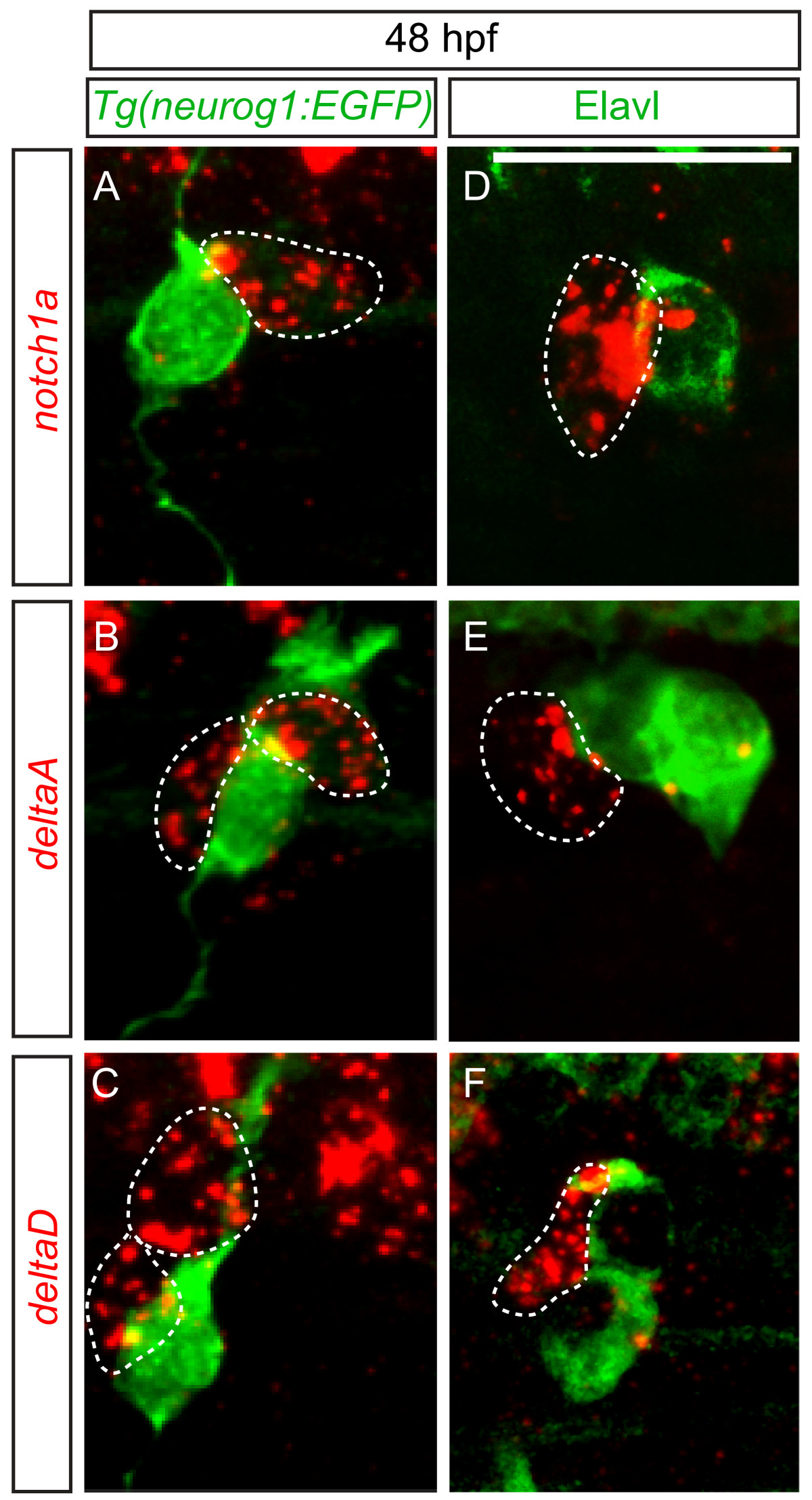
Fig. 5 delta and notch mRNAs are expressed in cells associated with dorsal root ganglia (DRG) neurons. (A-C) Confocal images of notch1a, deltaA and deltaD fluorescent RNA in situ hybridization in 48 hpf Tg(neurog1:EGFP) transgenic embryos. (A) Expression of notch1a (red, dashed line) is found in a cell that neighbors GFP + DRG neuron (green). Both deltaA (B) and deltaD (C) are expressed in cells surrounding GFP + neurons. (-F) Comparison of anti-Elavl labeling and notch1a, deltaA and deltaD RNA expression at 48 hpf. (D) A cell expressing notch1a (red) is seen adjacent to an Elavl-positive neuron (green). (E) deltaA is expressed in cells surrounding Elavl-positive neurons, as is deltaD (F). Scale bars, 20 μm. hpf, hours postfertilization.