
|
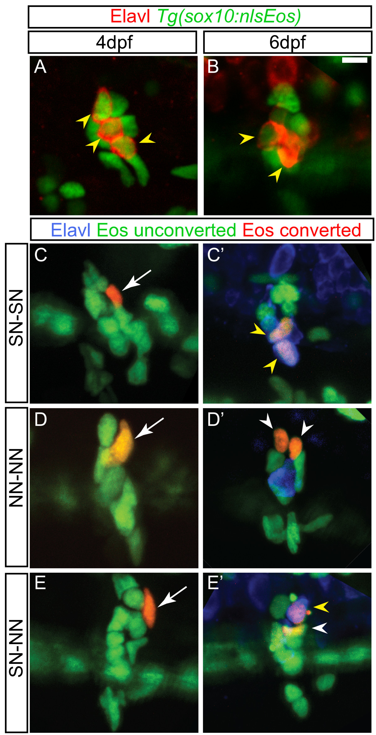
Fig. 4 Dividing progenitors within the larval dorsal root ganglia (DRG). (A-B) Confocal projections of ganglia-containing cells that express the Tg(sox10:nlsEos) transgene (green) and neurons labeled with Elavl (red). At four dpf (A) and six dpf (B) Tg(sox10:nlsEos) labels both neurons (yellow arrowheads) and non-neuronal cells. (C-E)Tg(sox10:nlsEos) cells were photoconverted by brief exposure to 405 nm light at four dpf, resulting in red fluorescence (white arrow). (C′-E′) At six dpf, larvae were labeled with Elavl (blue) and analyzed by confocal microscopy. Shown are examples of cells that divided and gave rise to two sensory neurons (SN-SN; yellow arrowheads) (C-C′), two non-neuronal cells (NN-NN; white arrowheads) (D-D′) or one sensory neuron and one non-neuronal cell (E-E′). Scale bar, 20 μm. dpf, days postfertilization.