
|
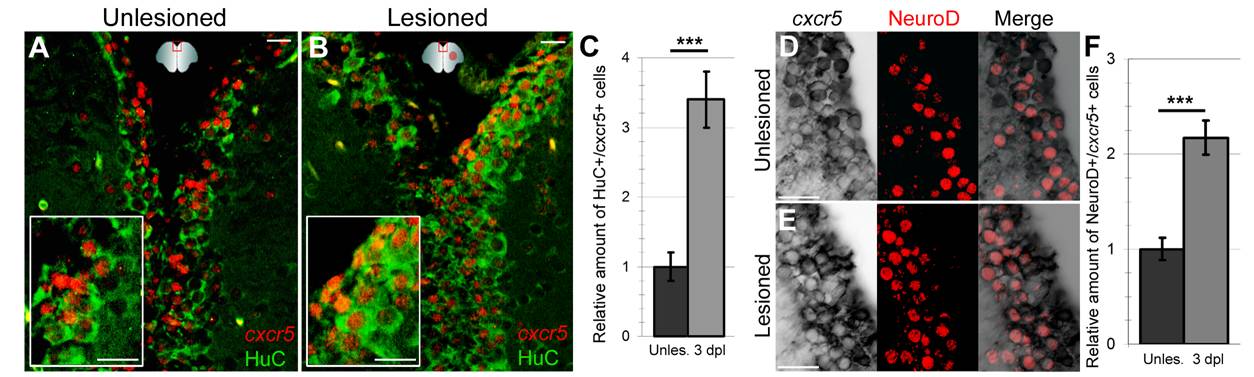
Fig. S2 Neurons express cxcr5. (A) cxcr5 fluorescent in situ hybridization (FISH) coupled to HuC immunohistochemistry (IHC) on a section of unlesioned adult zebrafish telencephalon. Inset is magnified image. (B)cxcr5 FISH coupled to HuC IHC on a section of 3 days post lesion (dpl) adult zebrafish telencephalon. Inset is magnified image. cxcr5 is expressed in neurons before and after injury. (C) Quantification graph for cxcr5-expressing HuC-positive cells. (D)cxcr5 chromogenic in situ hybridization (ISH) coupled to NeuroD IHC in unlesioned telencephalon. NeuroD-positive differentiating neurons, which are several cell diameters away from the ventricle express cxcr5 (white asterisks). cxcr5 expression is weaker in NeuroD-positive neurons in comparison to cxcr5-positive cells closer to the ventricle (yellow asterisks). In a transition zone between strong NeuroD-positive (white asterisks) and NeuroD-negative cells (yellow asterisks), cells express NeuroD and cxcr5 weakly (blue asterisks). (E)cxcr5 chromogenic ISH coupled to NeuroD IHC in 3 dpl telencephalon. NeuroD-positive differentiating neurons are more numerous, dispersed inside the parenchyma distantly in comparison to unlesioned telencephalons and express cxcr5 (white asterisks). cxcr5 expression is weaker in NeuroD-positive neurons in comparison to cxcr5-positive cells closer to the ventricle (yellow asterisks). (F) Quantification graph for cxcr5-positive NeuroD-expressing cells. The number of NeuroD/cxcr5 double-positive cells increase upon injury in adult zebrafish telencephalon. Scale bars 25 μm; n = 4 telencephalons for every set of analyses.