
Image
|
Figure Caption
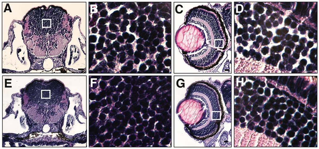
Fig. 4 Lama2cl501/cl501 fish exhibit growth abnormalities.
(A & E) Histological analysis of cross-section at 7 dpf revealed smaller brain in the mutant fish in comparison to wild-type fish. (B & F) Magnified views of brains showing tightly clumped cells in the mutant brains. (C & G) Wild-type as well as mutant fish displayed well-organized cellular layers in eyes. (D & H) Magnified views of the ganglion cell layer showed tightly organized cells with reduced extracellular space between the cellular layers in mutant fish in comparison to the wild-type.
Figure Data
Acknowledgments
This image is the copyrighted work of the attributed author or publisher, and
ZFIN has permission only to display this image to its users.
Additional permissions should be obtained from the applicable author or publisher of the image.
Full text @ PLoS One