
|
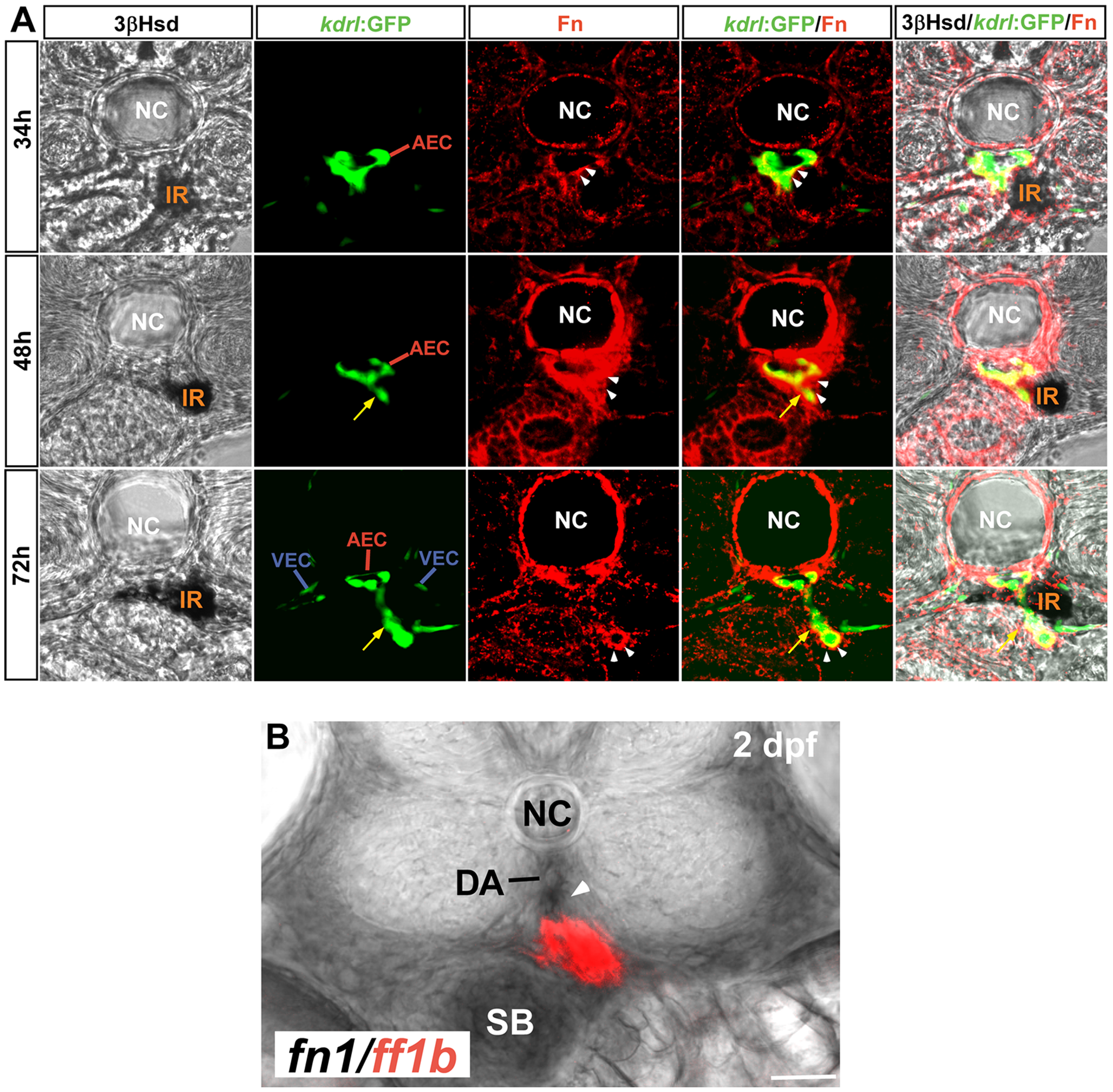
Fig. 1
The formation of IRV by angiogenic sprouting from the DA.
(A) Transverse sections of Tg(kdrl:GFP)s843 embryos at 34, 48 and 72 hpf visualized for 3β-Hsd activity (black), GFP (green) and Fibronectin (red). All sections are oriented with the posterior end toward top of page. While the IRV sprouting from the DA (yellow arrows) invades the interrenal tissue, steroidogenic interrenal cells form a protruding extension which migrates toward and across the central midline. Fibronectin (white arrowheads) accumulates at the interface between the endothelium and the interrenal tissue, and around the tip of growing IRV endothelium. (B) fn1 transcripts (black; indicated by white arrowhead) were detected around and ventral to the DA on the transverse section at the level of ff1b-expressing interrenal tissue (red) in a 2 dpf embryo. Abbreviations: notochord (NC), interrenal tissue (IR), arterial endothelial cell (AEC), venous endothelial cell (VEC), dorsal aorta (DA), swim bladder (SB). Scale bar is 50 μM.