
|
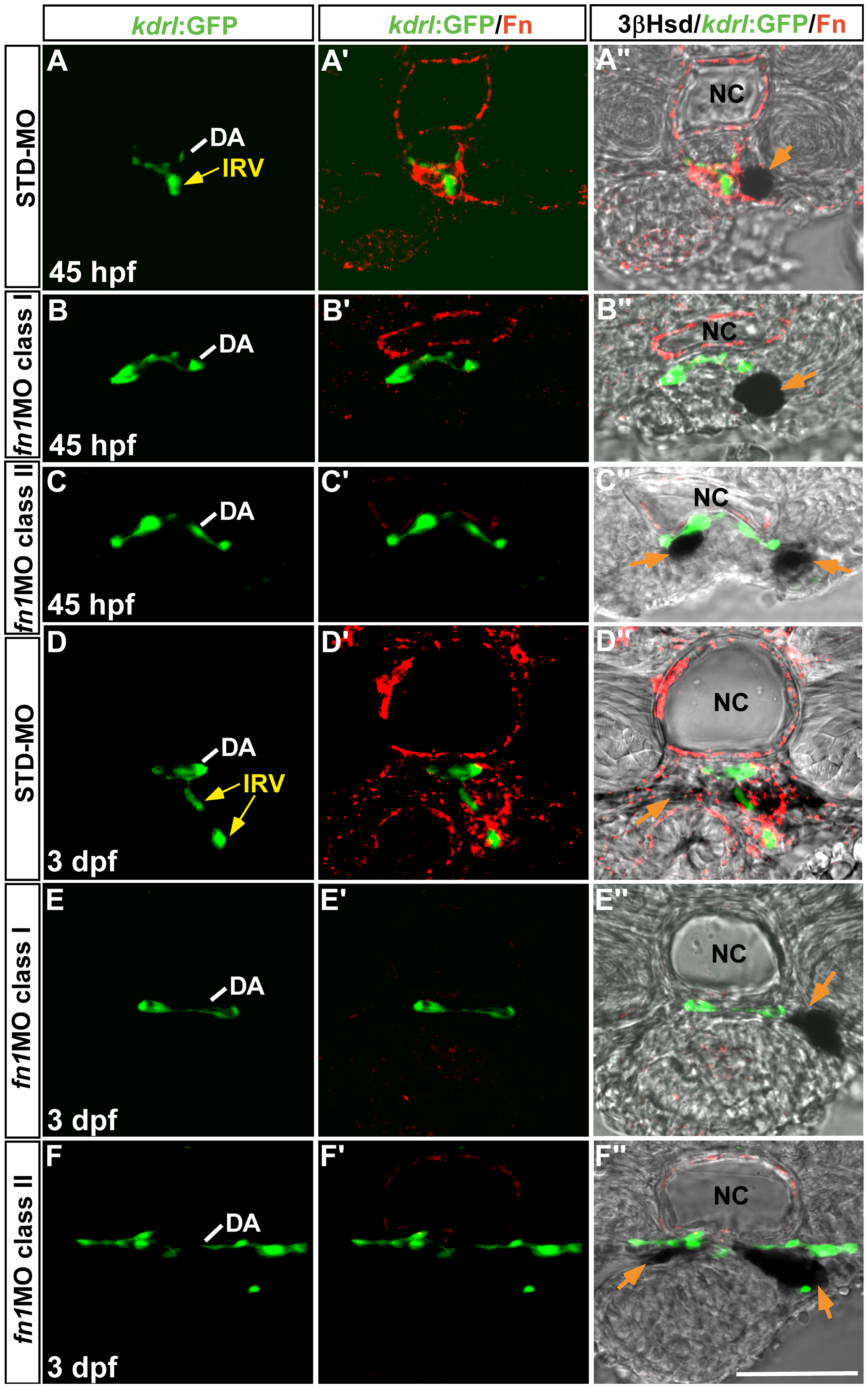
Fig. 3
The effect of fn1 knockdown on the interrenal tissue and the peri-interrenal vasculature.
Transverse sections of Tg(kdrl:GFP)s843 embryos injected with either STD-MO (A-A′′, D-D′′) or fn1MO (B-B′′, C-C′′, E-E′′, F-F′′) were assayed for 3β-Hsd activity (black), GFP (green) and Fn expression (red). All sections are oriented with the posterior end toward top of page. The IRV structure of a 3 dpf control embryo (D′-D′′) appeared to be discontinuous on a single confocal section, because the IRV at this stage extended slightly anteriorward and then posteriorward, after spanning ventrally through the interrenal tissue and before connecting to the AMA segment (Video S1). The formation of IRV is defective in the absence of peri-vascular Fn. Yellow arrows indicate the IRV sprouting from the DA. Orange arrow denotes the interrenal tissue. Abbreviations: dorsal aorta (DA), interrenal vessel (IRV), notochord (NC). Scale bar is 50 μM.