
Fig. 4
|
Fig. 4
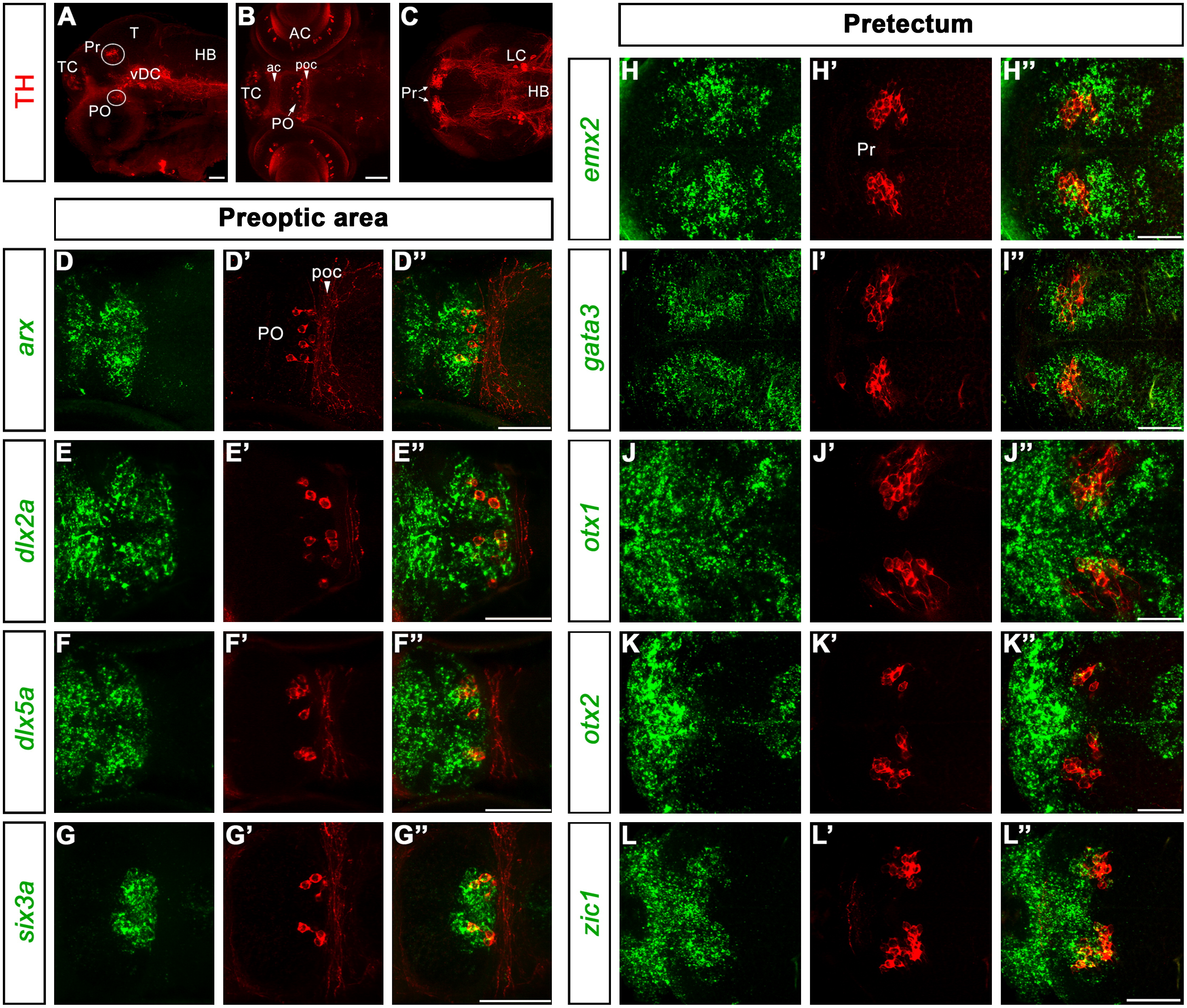
Expression of transcription factors characterizing DA neurons of the preoptic area and the pretectum. A: lateral overview is like in Fig. 1A. Here, the pretectal (Pr) and preoptic (PO) groups are framed by circles. B and C are dorsal z-projections encompassing the optical planes through the PO area (B, 34 μm projection) and the pretectum (C, 25 μm projection). D–G3 and H–L3 are higher magnified dorsal views of the PO (7–11 μm projections) and Pr (7–9 μm projections), respectively, at 96 hpf. The analyzed genes are indicated at the left side of each row. Abbreviations: ac, anterior commissure; AC, amacrine cells of the retina; HB, hindbrain; LC, locus coeruleus; PO, preoptic group; poc, postoptic commissure; Pr, pretectal group; T, tectum; TC, telencephalic cluster; vDC, ventral diencephalic cluster. Anterior is to the left. Scale bars: 50 μm.
Reprinted from Developmental Biology, 369(1), Filippi, A., Jainok, C., and Driever, W., Analysis of transcriptional codes for zebrafish dopaminergic neurons reveals essential functions of Arx and Isl1 in prethalamic dopaminergic neuron development, 133-149, Copyright (2012) with permission from Elsevier. Full text @ Dev. Biol.