
Image
|
Figure Caption
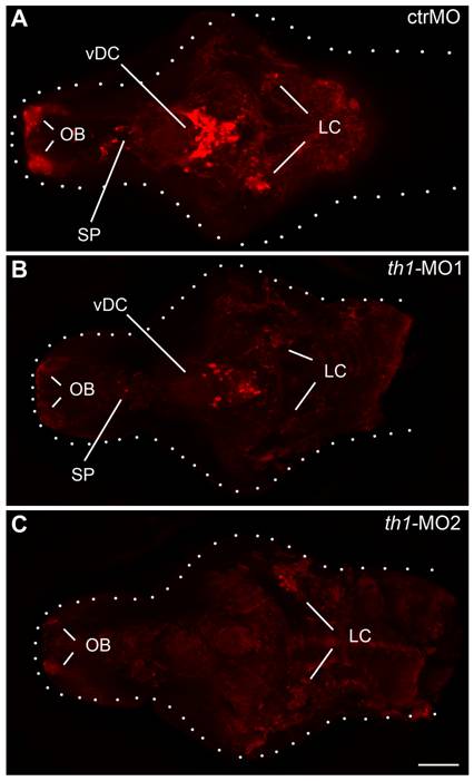
Fig. 3 Altered patterning of TH-positive cells in the larval brain of th1 morphants.
Immunofluorescence of TH-containing cells in the brains of control MO (A), th1-MO1 (B) and th1-MO2 (C) injected embryos (6 dpf). Confocal z-projections of larval brains are shown from a dorsal perspective, anterior to the left. OB olfactory bulb, LC locus coeruleus, SP subpallium, vDC ventral diencephalon. Dotted lines indicate brain outline. Scale bar = 100 μm.
Figure Data
Acknowledgments
This image is the copyrighted work of the attributed author or publisher, and
ZFIN has permission only to display this image to its users.
Additional permissions should be obtained from the applicable author or publisher of the image.
Full text @ PLoS One