
|
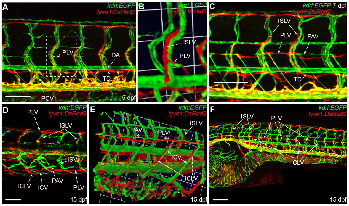
Fig. 3 The lateral lymphatics sprout from the intersegmental lymphatic vessel and form along the parachordal vessel and the intercostal vessel. All the images used in this figure are generated from the lyve1:DsRed2;kdrl:EGFP transgenic. (A) Lateral image of the trunk vessels at 5 dpf. (B) 3D reconstruction of the white box in A showing the initial sprout of the PLV originating from the ISLV. (C) Lateral image of the trunk vessels at 7 dpf, showing fusion of the PLV fragments. (D) Dorsal image of the trunk vessels at 15 dpf. (E) 3D reconstruction of the trunk vessels at 15 dpf, showing the dorsal migration of the ICLVs along the ICV. (F) Lateral image of the lateral lymphatics at 15 dpf. F is a montage image of eight z series stacks. DA, dorsal aorta; ICV, intercostal vessel; ICLV, intercostal lymphatic vessel; ISLV, intersegmental lymphatic vessel; ISV, intersegmental vessel; PAV, parachordal vessel; PCV, posterior cardinal vein; PLV, parachordal lymphatic vessel; TD, thoracic duct. Scale bars: 100 μm in A,C,D; 200 μm in F.