
Image
|
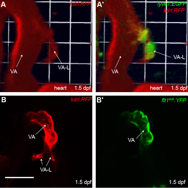
Figure Caption
Fig. S5 The ventral aorta lymphangioblast is derived from a non-arterial vessel near the ventral aorta. (A,A′) 3D reconstruction of the region where VA-L originates in the lyve1:EGFP;kdrl:RFP transgenic at 1.5 dpf. The VA-L co-expresses kdrl indicating its vascular origins. (B,B′) Ventrolateral images of the kdrl:RFP;flt1enh:YFP transgenic at 1.5dpf. The VA-L does not have flt1enh:YFP expression (B′) indicating its non-arterial origin (n=13). VA, ventral aorta; VA-L, ventral aorta lymphangioblast. Scale bars: 50 μm.
Acknowledgments
This image is the copyrighted work of the attributed author or publisher, and
ZFIN has permission only to display this image to its users.
Additional permissions should be obtained from the applicable author or publisher of the image.
Full text @ Development