
|
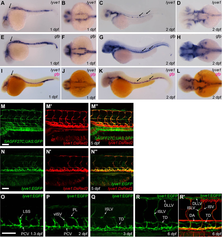
Fig. S1 The lyve1 promoter marks regions of endogenous lyve1 expression and labelled trunk lymphatics develop in a pattern known to occur in the emergence of the thoracic duct. (A-L) lyve1 probe (A-D), gfp probe (E-H) and lyve1 (BM Purple) gfp (Fast Red) probes (I-L) applied to lyve1:EGFP embryos at 1 dpf (A,B,E,F,I,J) and 2 dpf (C,D,G,H,K,L) (A,C,E,G,I,K, lateral view; B,D,F,H,J,L dorsal view of anterior region). Black arrows indicate areas where gfp expression does not match endogenous lyve1 expression. (M-M′′) Lateral image of a lyve1:DsRed2;SAGFF27C;UAS:YFP transgenic at 5 dpf showing SAGFF27C;UAS:YFP expression (M), lyve1:DsRed2 (M′) and overlapping lyve1:DsRed2;SAGFF27C;UAS:YFP expression in the trunk lymphatic vessels (M′′). (N-N′′) Lateral image of a lyve1:EGFP;lyve1:DsRed2 transgenic at 5 dpf showing lyve1:EGFP expression (N), lyve1:DsRed2 (N′) and overlapping lyve1:EGFP;lyve1:DsRed2 expression in the trunk lymphatic vessels and PCV (N′′). (O-Q) Lateral images of the developing trunk lymphatics in the lyve1:EGFP transgenic at 1.3 dpf (O), 2 dpf (P) and 3 dpf (Q). (R,R′) Lateral image of the developed trunk lymphatics in lyve1:EGFP transgenic at 6 dpf (R). Microangiography confirms that these vessels contain no blood-flow (R′). DA, dorsal aorta; DLLV, dorsal longitudinal lymphatic vessel; ISLV, intersegmental lymphatic vessel; ISV, intersegmental vessel; LSS, lymphatic secondary sprout; PCV, posterior cardinal vein; PL, parachordal lymphangioblasts; TD, thoracic duct; vISV, venous intersegmental vessel. Scale bar: 100 μm.