
|
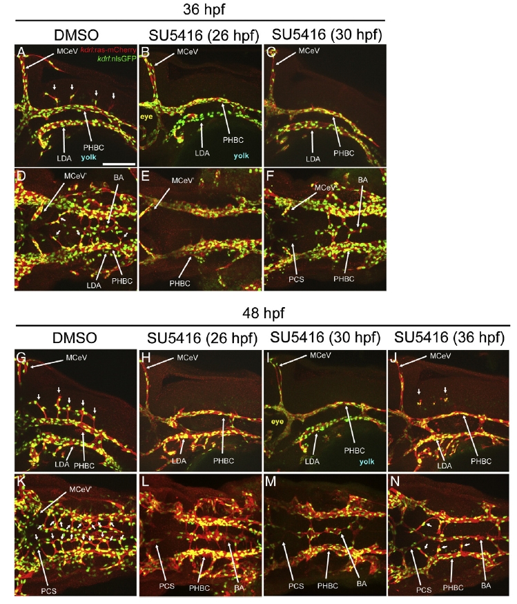
Fig. 6 SU5416 treatments reveal stage-specific VEGF signaling requirements for BA and CtA development. A-N, Maximum intensity confocal projections of immuno-fluorescently stained embryos carrying the endothelial reporters Tg(kdrl:ras-mCherry)s896 and Tg(kdrl:nls-GFP). Endothelium, red. Endothelial cell nuclei, green. Images show 36 (A-F) and 48 hpf (G-N) embryos treated with 0.01% of the drug vehicle DMSO (control) or with 1 μMof the VEGF signaling inhibitor SU5416 (in 0.01% DMSO) at the ages indicated in parentheses (26, 30 or 36 hpf). Abbreviations (see Table 1): vasculature, white (apostrophe, right side). Small white arrows, CtAs. A-C, G-J, Left lateral views. Anterior, left. Dorsal, up. D-F, K-N, Dorsal views. Anterior, left. Left side, bottom. Scale bar (A), 100 μm.
Reprinted from Developmental Biology, 357(1), Ulrich, F., Ma, L.H., Baker, R.G., and Torres-Vazquez, J., Neurovascular development in the embryonic zebrafish hindbrain, 134-51, Copyright (2011) with permission from Elsevier. Full text @ Dev. Biol.