
|
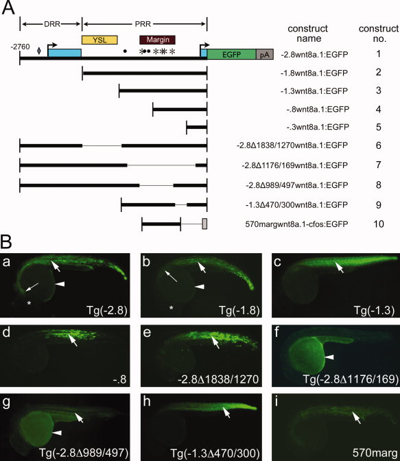
Fig. 2
Dissection of wnt8a cis-regulatory regions. A: Schematic diagram of reporter constructs used in this study, drawn to scale. Construct 1 (-2.8wnt8a.1:EGFP) contains the fragment extending 2,760 bp upstream of the wnt8a.1 translation initiation codon (-2,760). Salient functional features are indicated on construct 1. Light blue boxes, exons; arrows, transcription start sites. The diamond, asterisks, and dots indicate Zbtb4, FoxH1, and Ntl consensus binding sequences, respectively. Yellow box, YSL enhancer; maroon box, margin enhancer; DRR, distal regulatory region; PRR, proximal regulatory region. Thick bars indicate the regulatory region included in each construct. Thin lines indicate deleted regions. The grey box in construct 10 indicates the c-fos minimal promoter. B: EGFP reporter expression in stable or transient assays at 24 hpf. All images: lateral views, anterior to left. Reporter construct is indicated in lower right. a,b,c,f,g,h: Stable transgenic embryos. d,e,i: Transient expression assays. Arrows, somitic expression; arrowheads, YSL expression. Note that YSL fluorescence in stable lines is dimmer than that observed in transient assays, likely owing to stable integration of single-copy transposons. At 24 hpf, YSL fluorescence in stable lines is observed as an outline of the yolk that is slightly brighter than yolk autofluorescence. a,b: Long arrows, anterior limit of reporter fluorescence; asterisks, position of the eye.