
|
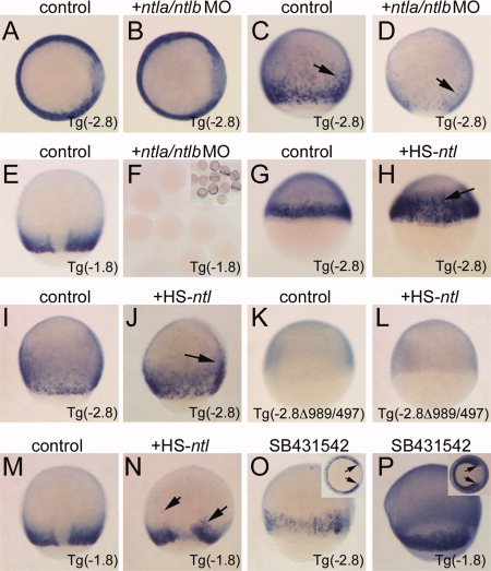
Fig. 6
Phase II expression requires Ntl stimulation of the margin enhancer. All images are in situ hybridizations for EGFP transcripts. Embryo genotypes are indicated in the lower right corner. Treatment is indicated above each panel. A,B: Shield stage, animal pole view, dorsal right. Note expression is unchanged in B compared to control. C,D: Eighty percent epiboly stage, lateral views, dorsal right. Note absence of expression in the margin in D; only scattered staining cells are observed (arrows). E,F: Eighty percent epiboly, dorsal view, anterior up. No staining is observed in MO-injected embryos bearing the PRR-only reporter (F; inset shows sibling uninjected embryos). G,H: Shield stage, lateral views, dorsal right. Ectopic ntla is sufficient to induce the Tg(-2.8) reporter at an early stage (H, arrow). I,J: Eighty percent epiboly, lateral view, dorsal right. Ectopic ntla is sufficient to induce the Tg(-2.8) reporter in the late gastrula (J, arrow). K,L: Shield stage, lateral view, dorsal right. Ectopic ntla cannot activate the reporter lacking the margin enhancer (L). M,N: Eighty percent epiboly, dorsal view, anterior up. Ectopic ntla can weakly activate PRR-only reporters in the late gastrula (N, arrows). O,P: Eighty percent epiboly, lateral views, dorsal right. Insets: vegetal views; arrows indicate expanded dorsal clearing indicative of Nodal inhibition. Tg(-2.8) embryos show phase II reporter expression (O), and PRR-only reporters are not inhibited by Nodal antagonism (P).