
|
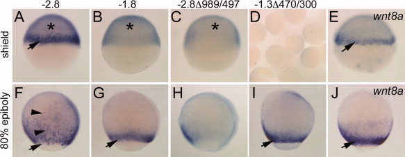
Fig. 4
Phase I and Phase II wnt8a reporter expression. All images are in situ hybridizations for EGFP transcripts in embryos from stable transgenic lines, except E, J, which show wnt8a expression in wild-type. Lateral view, dorsal right. A–E: Shield stage. F–J: Eighty percent epiboly. Reporter transgene is indicated above each row. Arrows, expression in the margin; asterisks, YSL expression, which at this stage manifests as a general blue stain underneath the epiblast layer of the embryo. YSL staining was confirmed by YSL fluorescence observed in live embryos. Representative group of embryos is shown in D. Arrowheads in F indicate scattered ingressing cells. Note qualitative difference between pattern in F from that in G and I and the wnt8a pattern in J.