
|
Fig. S1
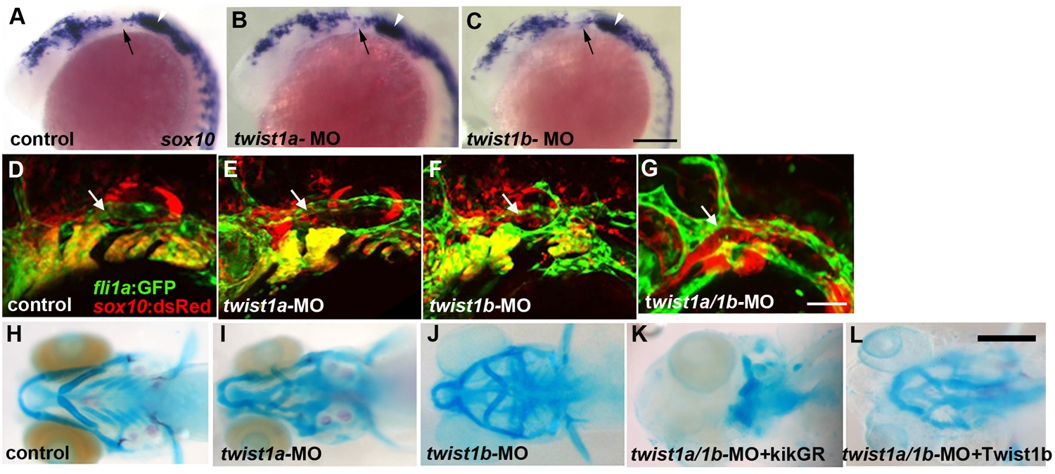
Twist1a and Twist1b function redundantly to specify ectomesenchyme. (A–C) In situs at 18 hpf show sox10 expression in un-injected, twist1a-MO, and twist1b-MO embryos. A few ectopic sox10-positive cells are seen in the second arches (arrows) of twist1a-MO and twist1b-MO embryos. White arrowheads denote the developing ear. (D–G) Confocal projections of fli1a:GFP; sox10:dsRed doubly transgenic embryos at 28 hpf show normal fli1a:GFP expression in un-injected control, twist1a-MO, and twist1b-MO embryos and loss of fli1a:GFP arch expression in twist1a/1b-MO embryos. Arrows indicate fli1a:GFP vascular expression which is unaffected in twist1a/1b-MO embryos. (H–L) Skeletal staining shows malformed mandibular and hyoid skeletons in twist1a-MO and twist1b-MO embryos compared to un-injected controls. In addition, co-injection of a Twist1b mRNA not targeted by the MOs partially rescued the head skeleton of twist1a/1b-MO embryos (n = 24/24), whereas co-injection of a control kikGR mRNA never rescued (n = 0/11). Scale bars = 50 μm.