
Image
|
Figure Caption
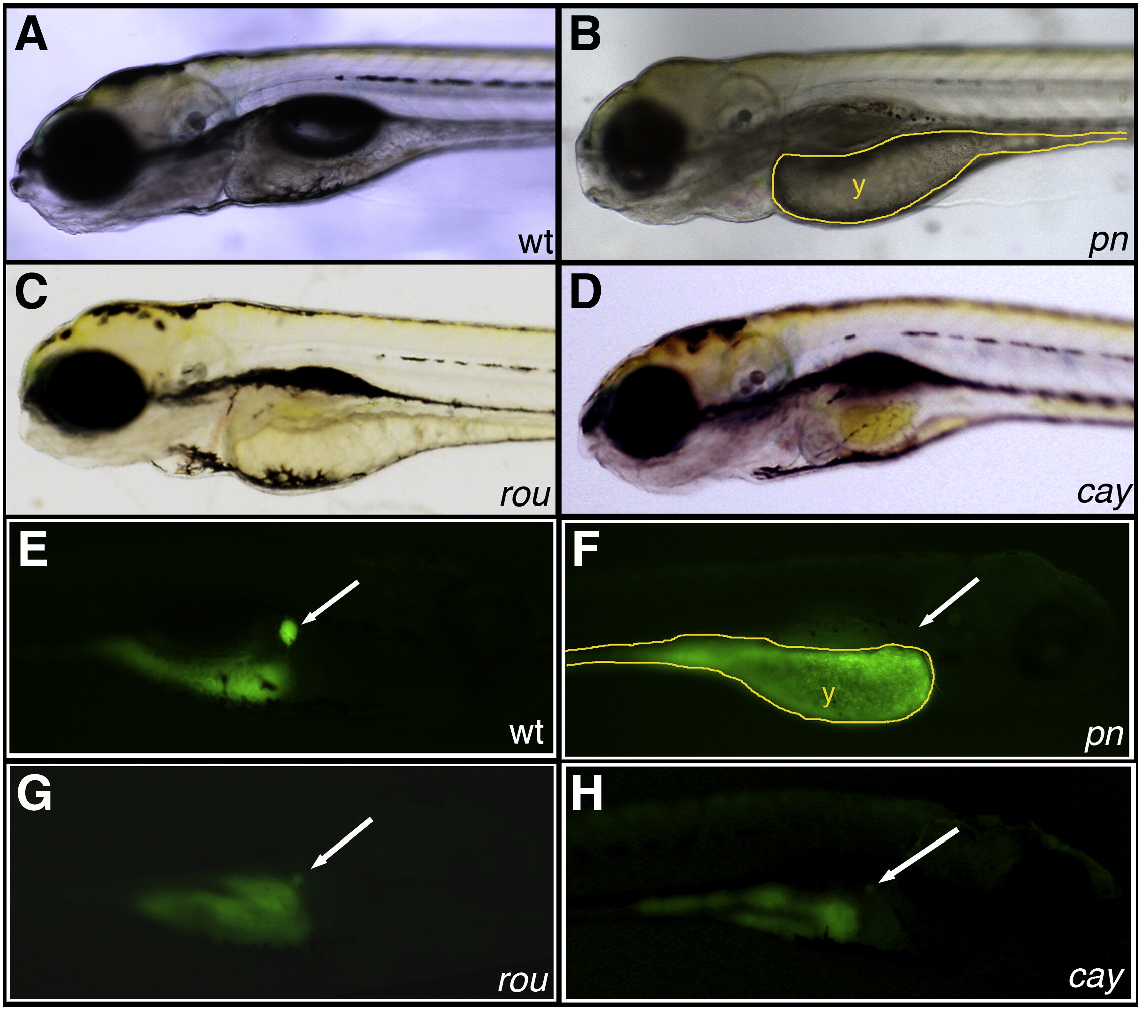
Fig. 1
Novel mutants with abnormal PED-6 uptake. (A–D) Left lateral views of live 5 dpf wild-type (A, wt), pekin (B, pn), rouen (C, rou), and cayuga (D, cay) larvae. pn demonstrates global hypopigmentation, underutilized yolk (y), absent swim bladder and some brain edema. (E–H) Right lateral views of the same 5 dpf larvae after PED-6 uptake, demonstrating normal intensity of the gallbladder in wt (E, white arrow), but absent gallbladders in the mutants (F–H, white arrows).
Figure Data
Acknowledgments
This image is the copyrighted work of the attributed author or publisher, and
ZFIN has permission only to display this image to its users.
Additional permissions should be obtained from the applicable author or publisher of the image.
Reprinted from Developmental Biology, 365(2), Eauclaire, S.F., Cui, S., Ma, L., Matous, J., Marlow, F.L., Gupta, T., Burgess, H.A., Abrams, E.W., Kapp, L.D., Granato, M., Mullins, M.C., and Matthews, R.P., Mutations in vacuolar H(+)-ATPase subunits lead to biliary developmental defects in zebrafish, 434-444, Copyright (2012) with permission from Elsevier. Full text @ Dev. Biol.