
Image
|
Figure Caption
Fig. 7
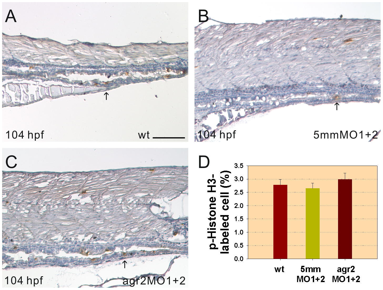
Intestinal cell proliferation is not altered in agr2 morphants.
Images of p-Histone H3-stained cells in the mid-intestines and posterior intestines of 104-hpf wild type embryos (A), agr2–5 mmMO1 and 5 mmMO2-coinjected embryos (B) and agr2 morphants (C) are shown. Arrows indicate p-Histone H3-stained cells. (D) Comparison of the percentages of p-Histone H3-stained M phase cells among wild type embryos, agr2–5 mmMO1 and 5 mmMO2-coinjected embryos, and agr2 morphants is shown. Error bars indicate the standard error. Scale bars represent 100 μm.
Figure Data
Acknowledgments
This image is the copyrighted work of the attributed author or publisher, and
ZFIN has permission only to display this image to its users.
Additional permissions should be obtained from the applicable author or publisher of the image.
Full text @ PLoS One