
|
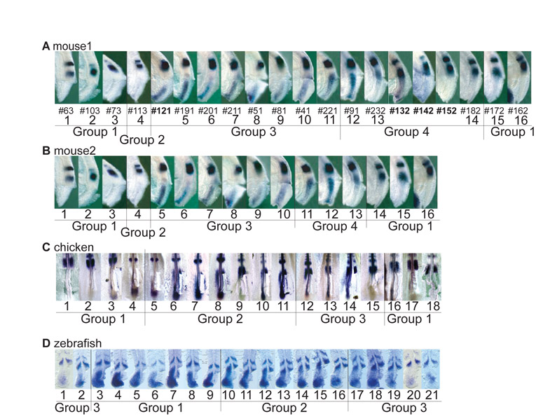
Fig. S1 Embryo assignment to local time windows for permutation analysis. It is often difficult, based on the in situ pattern, to decide whether an embryo oscillation phase is more advanced compared with that of another embryo that shows a closely related staining. We therefore divided the microarray series into groups, within which the relative position of embryos was hard to determine, thereby defining local time windows. Images of the embryos used in the microarray experiments and the groups corresponding to the local time windows used for the permutation analysis are shown for (A) mouse1, (B) mouse2, (C) chicken and (D) zebrafish.