
Image
|
Figure Caption
Fig. 9
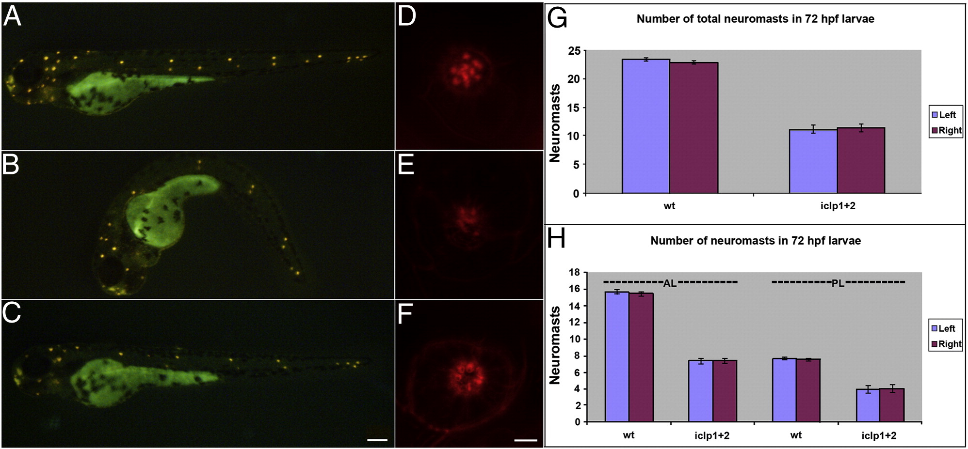
Development of lateral line (LL) in iclp morphants. (A–C) 4-Di-2-ASP staining of 72-hpf control (A), iclp morphant (B), and iclp morphant with capped RNAs (C). (D–F) Phalloidin staining of posterior LL neuromast at 5 dpf in control (D), iclp MOs-injected (E), and iclp MOs plus RNA-injected (F) larvae. Scale bar: 100 μm for (A–C), 5 μm for (D–E). (G, H) Comparison of numbers of neuromast stained with 4-Di-2-ASP in control larvae and iclp morphants. Wt (n = 10), MO (n = 15).
Figure Data
Acknowledgments
This image is the copyrighted work of the attributed author or publisher, and
ZFIN has permission only to display this image to its users.
Additional permissions should be obtained from the applicable author or publisher of the image.
Reprinted from Developmental Biology, 363(1), Shen, Y.C., Thompson, D.L., Kuah, M.K., Wong, K.L., Wu, K.L., Linn, S.A., Jewett, E.M., Shu-Chien, A.C., and Barald, K.F., The cytokine macrophage migration inhibitory factor (MIF) acts as a neurotrophin in the developing inner ear of the zebrafish, Danio rerio, 84-94, Copyright (2012) with permission from Elsevier. Full text @ Dev. Biol.