
|
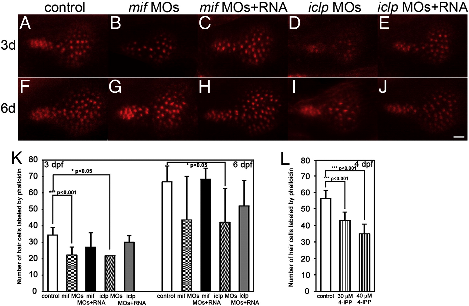
Fig. 6
Phalloidin staining of the sensory hair patch showed defects in mif and iclp morphants. (A–J) Phalloidin staining of saccular macula with 3d (A–E) and 6d (F–J) larvae. Anterior is to the left. (K) Numbers of HC labeled by phalloidin at 3 dpf (n = 9 for control, 10 for mif morphants, 13 for mif morphants with RNA rescue, 2 for iclp morphants, 6 for iclp morphants with capped RNA) and 6 dpf (n = 4 for control, 7 for mif morphants, 3 for mif morphants with RNA rescue, 5 for iclp morphants, 6 for iclp morphants with capped RNA). (L) Numbers of HC labeled by phalloidin at 4 hpf (n = 14 for DMSO control, 9 for 30 μM 4-IPP, 12 for 40 μM 4-IPP treatment). Scale bar: 10 μm.
Reprinted from Developmental Biology, 363(1), Shen, Y.C., Thompson, D.L., Kuah, M.K., Wong, K.L., Wu, K.L., Linn, S.A., Jewett, E.M., Shu-Chien, A.C., and Barald, K.F., The cytokine macrophage migration inhibitory factor (MIF) acts as a neurotrophin in the developing inner ear of the zebrafish, Danio rerio, 84-94, Copyright (2012) with permission from Elsevier. Full text @ Dev. Biol.