
|
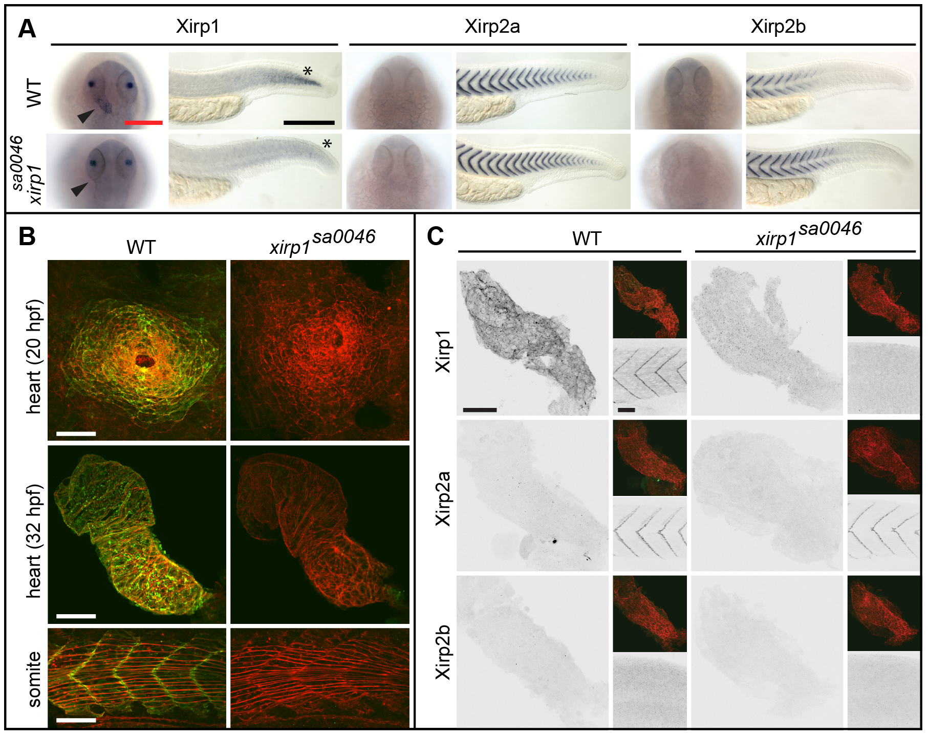
Fig. 6
Normal cardiogenesis and skeletal muscle development in the xirp1sa0046 mutant.
(A) Whole-mount in situ hybridization of xirp1, xirp2a and xirp2b on 24 hpf WT and xirp1sa0046 mutants reveal a loss of xirp1 expression in heart and muscle of xirp1sa0046 mutants. In contrast, neither xirp2a nor xirp2b expression are affected by lack of xirp1. In particular, neither xirp2a nor xirp2b are upregulated in the hearts of xirp1sa0046 mutants. Arrowheads indicate the position of the heart tube. Asterisks indicate the tip of the tail. Red scale bar: 200 μm, black scale bar: 250 μm. (B) Lack of xirp1 mRNA expression corresponds with the complete absence of Xirp1 (green) within the 20 hpf heart cone, 32 hpf heart tube, and 21 hpf skeletal muscle. The Actin counterstaining (red) demonstrates that cardiogenesis and skeletal muscle organization at these stages is comparable between WT and xirp1sa0046 mutants. Scale bars: 50 μm. (C) Immunohistochemical stainings for Xirp1, Xirp2a and Xirp2b on 24 hpf WT and xirp1sa0046 mutant hearts show complete absence of Xirp2a and Xirp2b. Top inserts depict overlay images of Actin (red) and Xirp1, Xirp2a or Xirp2b (green). Within the WT somitic muscle, both Xirp1 and Xirp2a are expressed whereas Xirp1 is absent in xirp1sa0046 mutant somitic tissue (bottom inserts). Scale bars: 50 μm.