
|
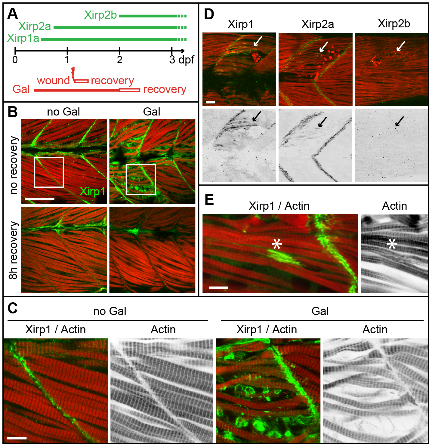
Fig. 1
Expression and localization of Xirps within Galanthamine- and laser-induced myocellular wounds.
(A) Schematic diagram summarizing the temporal order of Xirp expression within somitic muscle and in myocellular wounding assays. (B) Galanthamine (GAL) treatment between 80% epiboly and 2 dpf causes severe disruptions of somitic muscle organization and myofibrillar disarray (red: Actin) in 2 dpf zebrafish embryos. Notably, Xirp1 (green) is strongly expressed and localizes within cells most strongly disrupted by the treatment. These effects are completely reversible within several hours of recovery. Scale bar: 50 μm. (C) Details from inserts indicated in B (green: Xirp1; red: Actin). Scale bar: 10 μm. (D, E) Similarly, laser-induced muscle injury induces ectopic Xirp1 and Xirp2a localization to damaged myofibrils. In comparison, Xirp2b is not yet expressed at 33 hpf. Green: Xirp1, Xirp2a or Xirp2b; red: Actin. Arrows indicate the position of laser-induced injury within somitic tissue. Scale bars: 10 μm. Embryos were injured at 27 hpf (D) or 29 hpf (E) and fixed at 33 hpf (D) or 31.5 hpf (E), respectively.