
|
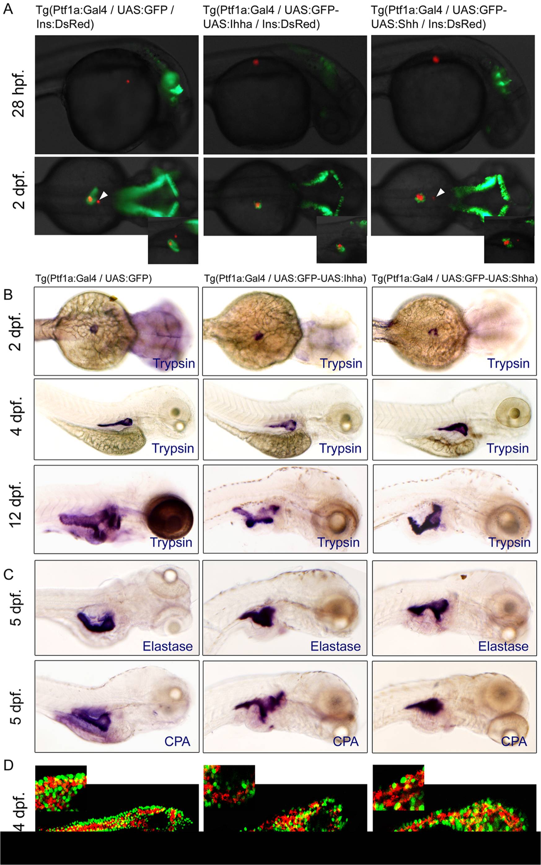
Fig. 2 Unaffected endocrine and exocrine differentiation by Hh over-expression.
(A) Fluorescence images showing the endocrine (RFP) and exocrine pancreas (GFP). When each transgenic fish is crossed with Ins-DsRed zebrafish, the development of insulin-expressing endocrine pancreas is not impaired by Hh over-expression. A smaller dot-like insulin-positive structure (white arrowheads) which corresponds to the anterior endocrine cells is observed in approximately half of the control and Hh-expressing embryos. (B, C) Whole mount ISH for trypsin, elastase, and carboxypeptidase A (CPA) at different time points. Over-expression of Hh ligands does not compromise the exocrine differentiation of the zebrafish pancreas, as evidenced by the proper and timely expression of trypsin. Expression of the other exocrine markers is also unaffected by Hh over-expression. Hh over-expression, however, induces subtle morphologic changes of the exocrine pancreas, showing a short, slender, and tortuous posterior pancreas compared to those of controls, which is evident by ISH for exocrine markers at 4 and 5 dpf and exaggerated at 12 dpf. (D) Confocal images of immunofluorescence staining for CPA. Regardless of transgene (GFP) expression, most acinar cells express CPA, suggesting unaffected exocrine differentiation by Hh over-expression. Lat., lateral.