
|
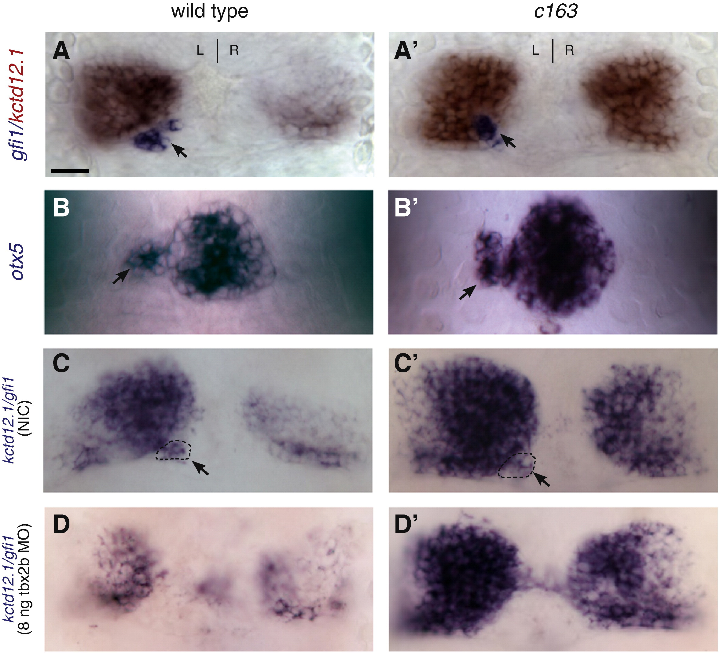
Fig. 3 The enlarged LsDh phenotype of sec61al1c163 is independent of the parapineal organ. (A) In wild-type larvae, the parapineal organ (gfi-1, blue) is found adjacent to the left LsDh (kctd12.1, red). (A′) Likewise, sec61al1c163 mutants possess a single parapineal organ on the left side of the brain. (B) The pineal complex (consisting of the pineal and parapineal, labeled by otx5) is similar in WT and (B′) sec61al1c163 mutant larvae. (C) Compared to an non-injected control (NIC), with a parapineal organ on the left side of the brain (dashed oval), (D) disruption of parapineal organ formation using a tbx2b antisense morpholino causes a reduction in the size of the left LsDh (kctd12.1). (C′, D′) However, in sec61al1c163 mutants, reduction of the parapineal organ has no effect on the size of the LsDh on either the left or right side. All views are dorsal. Panels A, C, D = 96 hpf; B = 72 hpf. Parapineal indicated by black arrowheads A–C′. Scale bar = 10 μm.
Reprinted from Developmental Biology, 360(1), Doll, C.A., Burkart, J.T., Hope, K.D., Halpern, M.E., and Gamse, J.T., Subnuclear development of the zebrafish habenular nuclei requires ER translocon function, 44-57, Copyright (2011) with permission from Elsevier. Full text @ Dev. Biol.