
|
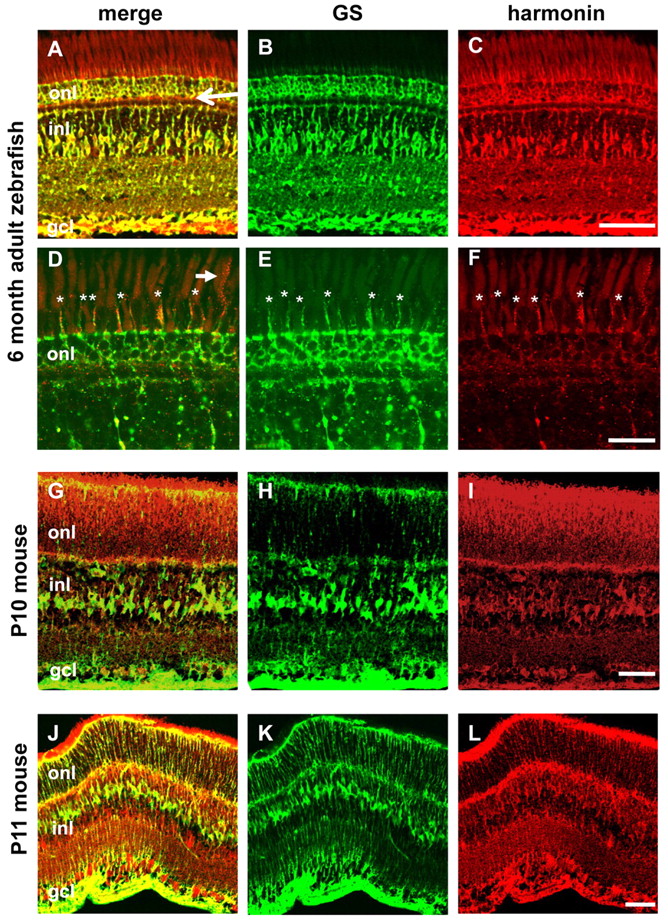
Fig. 6 Harmonin localizes in Müller cells through all zebrafish life stages and is evolutionarily conserved. (A–F) Localization of anti-zebrafish harmonin (red) in Müller cells persists in adult zebrafish retinas. Some low level signal is sometimes observed in photoreceptor synapses (arrow in A) and in photoreceptor outer segments (arrow in D). The strongest signal within the photoreceptor layer is associated with Müller cell processes projecting into the subapical region (asterisks). Nonspecific fluorescence of photoreceptor outer segments at this stage can be compared with panel H in supplementary material Fig. S4. (G–L) Anti-mouse harmonin (SDI; G,I) and anti-human harmonin (Santa Cruz; J,L) detect localization in photoreceptors and Müller cells [glutamine synthetase (GS) is labeled in green; harmonin antibodies in red] in retinas from P10–P11 mouse pups. The outer nuclear layer (onl), inner nuclear layer (inl) and ganglion cell layer (gcl) are labeled in merged panels. Scale bars: 50 μm (A–C,G–L); 20 μm (D–F).